ABSTRACT
- Three bacterial strains, W99T, S113T, and S268T, were isolated from marine algae and subjected to polyphasic taxonomic characterization. Phylogenetic and phylogenomic analyses placed W99T within the genus Shimia, whereas S113T and S268T formed two distinct genus-level lineages within the family Roseobacteraceae. The three isolates were Gram-stain-negative, non-motile, and obligately aerobic, with optimum growth at 25, 25, and 30°C, respectively, and at 2.0% (w/v) NaCl. Ubiquinone-10 was the sole respiratory quinone in all three strains. The major fatty acids were 11-methyl C18:1 ω7c and summed feature 8 (C18:1 ω7c and/or C18:1 ω6c) in strain W99T; C16:0 and summed feature 8 in strain S113T; and 11-methyl C18:1 ω7c, cyclo-C19:0 ω8c, and summed feature 8 in strain S268T. The major polar lipids were phosphatidylglycerol and phosphatidylcholine, with diphosphatidylglycerol additionally detected in strain S268T. Polyphasic taxonomic analyses demonstrated that strain W99T represents a novel species of the genus Shimia, for which the name Shimia rhodophyticola sp. nov. is proposed (W99T = KACC 24154T = JCM 38196T), whereas strains S113T and S268T represent two novel genera and species, Neomarinovum algicola gen. nov., sp. nov. (S113T = KACC 23417T = JCM 38210T) and Phycorhabdus gracilariae gen. nov., sp. nov. (S268T = KACC 24156T = JCM 38211T), respectively. In addition, comprehensive phylogenomic and genome-based relatedness analyses support the taxonomic revision of nineteen taxa within the family Roseobacteraceae, providing a revised genome-based taxonomic framework that better reflects the evolutionary relationships within the family.
-
Keywords: Shimia rhodophyticola, Neomarinovum algicola, Phycorhabdus gracilariae, new taxa, reclassification, Roseobacteraceae, marine algae
Introduction
The family Roseobacteraceae Liang et al. 2021, recently separated from the family Rhodobacteraceae based on genome-scale phylogenetic and genotypic analyses, comprises a diverse group of marine bacteria with broad physiological and ecological diversity (Liang et al., 2021). As of July 2026, the family includes 144 validly published genera isolated from a wide range of marine habitats, including seawater (Hwang and Cho, 2008; Lanclos et al., 2025), tidal mudflats (Lee et al., 2020), marine solar salterns (Wang et al., 2023), sea sand (Thongphrom et al., 2017), phycospheres (Coe et al., 2023), and marine algae (Jung et al., 2019; Kim et al., 2025; Lee et al., 2024). Recent genome-based studies have substantially improved our understanding of evolutionary relationships within the family (Wirth and Whitman, 2018; Zhang et al., 2023), but the current classification of several genera remains incongruent with their phylogenomic placement.
The genus Shimia was established with Shimia marina as the type species (Choi and Cho, 2006) was subsequently expanded through taxonomic reclassifications, including the transfer of Thalassobius aestuarii (Yi and Chun, 2006) and Thalassobius abyssi (Nogi et al., 2016) to the genus Shimia by Wirth and Whitman (2018). The genus currently comprises 13 species with validly published names and one species whose name has not been validly published. Members of the genus have been recovered from diverse marine environments, including seawater (Arahal et al., 2019; Chen et al., 2023; Hameed et al., 2013; Kim et al., 2008), sediments (Nogi et al., 2015, 2016; Yi and Chun, 2006; Zhu et al., 2021), corals (Chen et al., 2011), and marine invertebrates (Hyun et al., 2013; Kim et al., 2014). They are characterized as Gram-stain-negative, aerobic, rod-shaped bacteria possessing ubiquinone-10 (Q-10) as the predominant respiratory quinone, with 11-methyl C18:1 ω7c and summed feature 8 (C18:1 ω7c and/or C18:1 ω6c) as the major fatty acids. The major polar lipids are typically phosphatidylglycerol (PG) and phosphatidylcholine (PC), whereas phosphatidylethanolamine (PE) and diphosphatidylglycerol (DPG) vary among species. However, recent genome-based studies have suggested that the current circumscription of Shimia, as well as several other genera within the family Roseobacteraceae, does not fully reflect their evolutionary relationships.
Three isolates, W99T, S113T, and S268T, were recovered from marine algal phycospheres during our investigation of algae-associated bacterial diversity (Bayburt et al., 2024, 2026; Kim et al., 2025). Their taxonomic positions were evaluated using phylogenetic, genomic, phenotypic, and chemotaxonomic evidence. Genome-scale comparisons of selected Roseobacteraceae taxa additionally identified several discrepancies between their current classifications and inferred evolutionary relationships. Accordingly, this study describes Shimia rhodophyticola sp. nov., Neomarinovum algicola gen. nov., sp. nov., and Phycorhabdus gracilariae gen. nov., sp. nov., and proposes the taxonomic revision of nineteen taxa within the family Roseobacteraceae. These taxonomic revisions provide a revised genome-based framework that better reflects the evolutionary relationships within the family Roseobacteraceae.
Materials and Methods
Bacterial isolation and cultivation
Strains W99T, S113T, and S268T were isolated from the phycospheres of marine algae collected from the Republic of Korea as previously described (Kim et al., 2025). Strain W99T was isolated from the marine red alga Schizymenia sp. collected at Guryepo Beach (36°53′30.6″ N, 126°11′52.8″ E), strain S113T from the marine green alga Chaetomorpha sp. collected at Nogu Port (34°45′17.0″ N, 128°03′20.9″ E), and strain S268T from the marine red alga Gracilaria sp. collected at Mulgeon Port (34°46′50.5″ N, 128°02′56.5″ E). In brief, loosely associated microorganisms were removed from the algal samples by washing with sterile artificial seawater (ASW; 20.0 g NaCl, 2.9 g MgSO4, 4.5 g MgCl2·6H2O, 0.6 g KCl, 1.8 g CaCl2·2H2O per liter). The washed samples were homogenized with a T10 basic homogenizer (IKA, Germany), and serial dilutions of the homogenates were prepared in sterile ASW. Each dilution (100 µl) was inoculated onto marine agar (MA; MBcell, Korea) and incubated aerobically at 25°C until colony formation. Colony differentiation was performed by restriction analysis of 16S rRNA gene amplicons. The genes were amplified with primers 27F and 1492R (Lane, 1991), followed by digestion of the PCR products with HaeIII and HhaI. Restriction profiles were resolved on 2% agarose gels, and amplicons representing distinct profiles were selected for sequencing with primer 340F (Kim et al., 2025). Sequencing was performed by Macrogen (Korea), and the resulting sequences were compared with those of type strains of validly published species using the EzBioCloud Identify service (Chalita et al., 2024). Based on these analyses, strains W99T, S113T, and S268T were selected for further taxonomic characterization. Routine cultivation was performed aerobically on MA or in marine broth (MB; MBcell) at 25°C for 3 days. For long-term preservation, cultures were stored at −80°C in MB supplemented with 15% (v/v) glycerol.
Phylogenetic analysis using 16S rRNA gene sequences
Nearly complete 16S rRNA gene sequences of strains W99T, S113T, and S268T were generated from PCR products amplified with primers 27F and 1492R. Additional sequence reads were obtained with primers 518R (5′-ATT ACC GCG GCT GCT GG-3′) and 805F (5′-GAT TAG ATA CCC TGG TAG TC-3′) (Kim et al., 2025), and the reads generated with 340F, 518R, and 805F were assembled to obtain the final sequences. Sequence similarities between the isolates and their closest type strains were determined using the EzBioCloud server (Chalita et al., 2024). For phylogenetic reconstruction, the 16S rRNA gene sequences were aligned with Infernal v1.1.4 using the Rfam RF00177 covariance model (Nawrocki and Eddy, 2013). Maximum-likelihood (ML), neighbor-joining (NJ), and maximum-parsimony (MP) analyses were independently conducted in MEGA11 (Tamura et al., 2021), and node support was assessed from 1,000 bootstrap replicates. Based on 16S rRNA gene sequence similarities and phylogenomic relationships, Shimia aestuarii KCTC 12049T, Thiosulfatihalobacter marinus KCTC 82723T, Marinovum algicola JCM 21266T, and Primorskyibacter aestuariivivens KCTC 52455T were selected as reference strains for comparative genomic, phenotypic, and chemotaxonomic analyses. The reference strains for phenotypic and chemotaxonomic comparisons were selected primarily based on their taxonomic relevance to strains W99T and S113T. For strain S268T, closely related taxa were included in the phylogenomic and genome-relatedness analyses to assess its taxonomic position, whereas additional strains were not included in the experimental phenotypic and chemotaxonomic comparisons because these analyses were intended to provide supplementary characterization rather than to determine its genus-level affiliation.
Ecological distribution analysis based on 16S rRNA gene sequences
To estimate the ecological distribution of strains W99T, S113T, and S268T, their 16S rRNA gene sequences were queried against 296,393 metagenomic 16S rRNA amplicon datasets representing diverse environments using the Integrated Microbial Next-Generation Sequencing (IMNGS) platform (Lagkouvardos et al., 2016) with a 99.0% sequence similarity threshold. The datasets included marine and freshwater environments (seawater, estuaries, marine sediments, and freshwater), marine hosts (algae, corals, sponges, crustaceans, molluscs, oysters, and sea anemones), beach sand, biofilms, and terrestrial and engineered environments, including soil, compost, plants, wastewater, and activated sludge.
Whole genome sequencing, genome-based phylogeny, and genome-relatedness analysis
Genomic DNA was extracted from strains W99T, S113T, and S268T using the Wizard Genomic DNA Purification Kit (Promega, USA) according to the manufacturer’s instructions. Libraries were prepared using the Ligation Sequencing Kit (SQK-LSK109) with Native Barcoding Expansions EXP-NBD104 and EXP-NBD114 (Oxford Nanopore Technologies, UK) and sequenced on a MinION platform using R9.4.1 chemistry. Basecalling was performed in MinKNOW using the corresponding R9.4.1 MinION model. Reads were adapter-trimmed with Porechop v0.2.4 and filtered with NanoFilt v2.8.0 using minimum thresholds of Q10 and 200 bp. De novo assembly of the filtered reads was performed with Flye v2.9.1 in nano-hq mode (Kolmogorov et al., 2019), specifying a 5 Mb genome size and 50× coverage. The assemblies underwent one Flye polishing cycle, with no subsequent polishing step. Mean genome coverage was obtained from the final Flye assembly statistics. Genome completeness and contamination were assessed using CheckM2 v1.0.2 (Chklovski et al., 2023), and genomes were annotated using the NCBI Prokaryotic Genome Annotation Pipeline (PGAP; software revision 6.11).
Phylogenomic analysis included 206 genomes representing members of the family Roseobacteraceae, selected based on the phylogenomic relationships reported by Liang et al. (2021), with emphasis on taxa closely related to the three isolates and those relevant to the proposed taxonomic revisions. Additional closely related genera were selected based on 16S rRNA gene sequence similarities, and all available type-strain genomes of validly published species within the selected genera were included. GTDB-Tk v2.4.1 with GTDB release 226 (Chaumeil et al., 2022) was used in the de novo workflow to identify and align the bac120 single-copy marker proteins and to generate the trimmed concatenated alignment. The canonical bacterial mask was applied, with missing markers represented as gaps. The resulting concatenated alignment was used to reconstruct an ML phylogenomic tree in MEGA11 (Tamura et al., 2021) with 1,000 bootstrap replicates.
Genome relatedness was quantified using OrthoANIu for average nucleotide identity (ANI) (Yoon et al., 2017), Genome-to-Genome Distance Calculator (GGDC 3.0) with formula 2 for digital DNA-DNA hybridization (dDDH) (Meier-Kolthoff et al., 2022), and EzAAI v1.2.3 for average amino acid identity (AAI) (Kim et al., 2021). Percentage of conserved proteins (POCP) values were obtained according to Qin et al. (2014). AAI matrices were hierarchically clustered and visualized with ComplexHeatmap v2.14.0 in R v4.2.3, whereas pairwise ANI, AAI, and POCP relationships among members of the family Roseobacteraceae were plotted using ggplot2 and ggpubr.
Phenotypic, physiological, and biochemical characterization
Physiological growth limits of strains W99T, S113T, and S268T were determined using MA or MB, as appropriate. Temperature tolerance was assessed on MA from 5 to 50°C at 5°C intervals, whereas pH tolerance was examined in MB after 3 days of incubation over the range pH 3.0–12.0 at 1.0-unit intervals using citrate (pH 3.0–5.0), phosphate (pH 6.0–8.0), or carbonate-bicarbonate (pH 9.0–12.0) buffers. NaCl tolerance was evaluated in laboratory-prepared MB containing 0–10°C (w/v) NaCl. Growth on different nutrient media was tested on MA, R2A agar, LB agar, TSA, and NA adjusted to approximately 2% (w/v) NaCl after incubation at 25°C for 3 days. Unless otherwise specified, subsequent characterization was performed at the optimum temperature of each strain (25°C for strains W99T and S113T and 30°C for strain S268T). The ability to grow anaerobically was tested on MA for 21 days at each strain’s optimum temperature using a GasPak Plus system (BBL, USA). Morphological characteristics and motility were observed by phase-contrast microscopy (Zeiss Axio Scope.A1; Carl Zeiss, Germany) using cells grown on MA for 3 days at the optimum temperature of each strain. Cell ultrastructure was further examined by transmission electron microscopy (JEM-F200; JEOL, Japan) after negative staining with UranyLess (Electron Microscopy Sciences, USA) for 1 min (Benmeradi et al., 2015). Gliding behavior was examined on MA solidified with 0.3% (w/v) agar following Bernardet et al. (2002), and Gram reactions were determined with a commercial staining kit (bioMérieux, France). Catalase and oxidase activities were determined by bubble production in 3% (v/v) H2O2 (Junsei, Japan) and oxidation of 1% (w/v) tetramethyl-p-phenylenediamine (Merck, USA), respectively (Smibert and Krieg, 1994). All reference strains were examined under the same experimental conditions as the three isolates to permit direct phenotypic comparison. Substrate-degrading activities toward esculin, casein, starch, tyrosine, Tween 20, and Tween 80 were examined on MA according to Smibert and Krieg (1994). Additional biochemical characteristics were determined using the API 20NE system (bioMérieux, France) with inocula prepared in ASW according to the manufacturer's instructions.
Chemotaxonomic characterization
Chemotaxonomic analyses were conducted with exponentially growing cultures. Quinones prepared from W99T, S113T, and S268T according to Minnikin et al. (1984) were separated by reversed-phase HPLC using an LC-20A system equipped with an SPD-M20A diode-array detector and a Kromasil column (250 × 4.6 mm). Methanol/isopropanol (2:1, v/v) served as the mobile phase at 1 ml/min. Fatty acid profiles of the isolates and reference strains were determined from exponential-phase MB cultures (OD600 = 0.7–0.8) grown at their respective optimum temperatures. Fatty acid methyl esters prepared by the standard MIDI procedure were analyzed by gas chromatography (Hewlett Packard 6890), and components were assigned using the RTSBA6 database of Sherlock MIS v6.0B (Sasser, 1990). Polar lipid extracts prepared from exponentially growing cultures were resolved by two-dimensional thin-layer chromatography (TLC) following Minnikin et al. (1984). Total polar lipids, aminolipids, phospholipids, and glycolipids were detected with 10% (w/v) ethanolic molybdophosphoric acid, ninhydrin, Dittmer-Lester reagent, and α-naphthol/sulfuric acid, respectively; authentic PG, PC, and DPG standards were used for confirmation.
Results and Discussion
Isolation and phylogenetic characteristics based on 16S rRNA gene sequences
Screening of bacteria recovered from marine algal phycospheres yielded three putatively novel Roseobacteraceae isolates, designated W99T, S113T, and S268T. Their nearly complete 16S rRNA gene sequences were 1,404, 1,435, and 1,403 bp, respectively. Strain W99T shared the highest 16S rRNA gene sequence similarity with S. aestuarii DSM 15283T (99.9%), followed by Shimia abyssi JAMH 043T (98.3%). Strain S113T was most closely related to Rhodalgimonas zhirmunskyi KMM 6723T (97.3%) and Shimia sediminis ZQ172T (97.2%), whereas strain S268T shared the highest sequence similarity with S. aestuarii DSM 15283T (96.2%) and Aestuariibius insulae DBTF-13T (96.2%). Based on the accepted 16S rRNA gene sequence similarity threshold for species delineation (98.5–98.7%) (Riesco and Trujillo, 2024), strains S113T and S268T are likely to represent novel species, whereas the taxonomic status of strain W99T could not be resolved based solely on 16S rRNA gene sequence analysis.
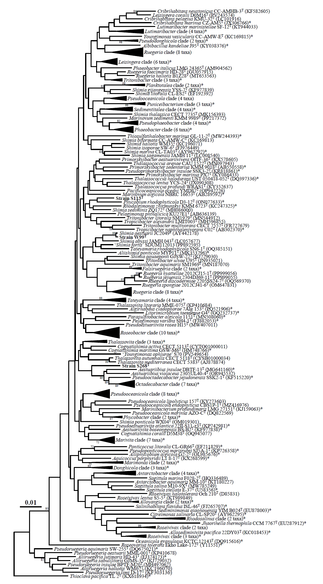
An NJ phylogenetic tree based on 16S rRNA gene sequences showed that strain W99T formed a robust cluster with S. aestuarii DSM 15283T (100% bootstrap support; Fig. 1). However, this clade, together with several other Shimia species, was clearly separated from the lineage containing the type species, Shimia marina. This topology was consistently recovered in the ML and MP trees (Fig. S1), indicating that 16S rRNA gene sequence-based phylogenies provide insufficient resolution to accurately resolve phylogenetic relationships within the genus Shimia. Strain S113T consistently clustered with Phycobium rhodophyticola D3-12T in the NJ, ML, and MP trees, but the bootstrap support was low in all cases, indicating that its phylogenetic position could not be resolved confidently. In contrast, strain S268T formed a distinct phylogenetic lineage in the NJ, ML, and MP trees, but its phylogenetic position relative to neighboring taxa remained unresolved. Collectively, these results demonstrate that the genus-level affiliations of strains W99T, S113T, and S268T could not be determined confidently based solely on 16S rRNA gene phylogeny, necessitating genome-based analyses to clarify their taxonomic positions.
Furthermore, the 16S rRNA gene phylogenies showed that several genera within the family Roseobacteraceae did not form distinct monophyletic clades but were interspersed throughout the trees. This pattern indicates that 16S rRNA gene sequences provide insufficient phylogenetic resolution for robust genus-level classification within the family and suggests that the taxonomy of several taxa should be reassessed using genome-based approaches.
Ecological distribution
Ecological distribution analysis using the IMNGS platform at a 99.0% 16S rRNA gene sequence similarity threshold showed that sequences closely related to strains W99T, S113T, and S268T were predominantly detected in marine environments (Table S1). Sequences closely related to W99T and S113T were frequently detected in sea anemone microbiomes (76.7% prevalence; average relative abundance [ARA], 3.08%) and the marine diatom Seminavis robusta (44.4% and 88.9% prevalence, respectively). Closely related sequences were also detected in various marine invertebrate-associated datasets, including those from the sea urchin Tripneustes gratilla, spiny lobster Panulirus ornatus, crab Cancer borealis, and corals, whereas sequences related to S268T generally showed lower prevalence. In contrast, sequences related to the three strains were rare or absent in terrestrial and anthropogenic environments, including compost, plants, and human-associated samples. Although the strains were isolated from marine algal phycospheres, the detection of closely related sequences across diverse marine host-associated habitats suggests that phylogenetically related bacteria may occur beyond algal hosts. Overall, these results suggest a predominantly marine distribution of 16S rRNA gene sequences closely related to the three strains.
Genome sequencing and genomic features
After quality filtering, read yields for strains W99T, S113T, and S268T were 335.8, 323.7, and 640.9 Mb, respectively, with corresponding read N50 values of 19,422, 11,491, and 28,075 bp. De novo assembly generated draft genomes of approximately 4,262, 4,314, and 3,864 kb, with average coverage depths of 76.0×, 73.0×, and 162.0× and assembly N50 values of 4,008,202, 4,022,761, and 3,667,163 bp, respectively. The genome-derived 16S rRNA gene sequences were identical to those obtained by PCR-based sequencing. CheckM2 indicated high estimated completeness and low estimated contamination for all three assemblies, with completeness and contamination values of 100.0% and 0.6% for W99T, 100.0% and 0.2% for S113T, and 93.2% and 1.2% for S268T, respectively (Chklovski et al., 2023).
The genomic DNA G + C contents of strains W99T, S113T, and S268T were 60.5, 60.0, and 56.1 mol%, respectively, consistent with those of their closely related taxa. Detailed genomic features, including genome size, G + C content, and numbers of coding sequences, tRNA genes, and rRNA genes, are summarized in Table S2.
Genome-based phylogeny and genome relatedness
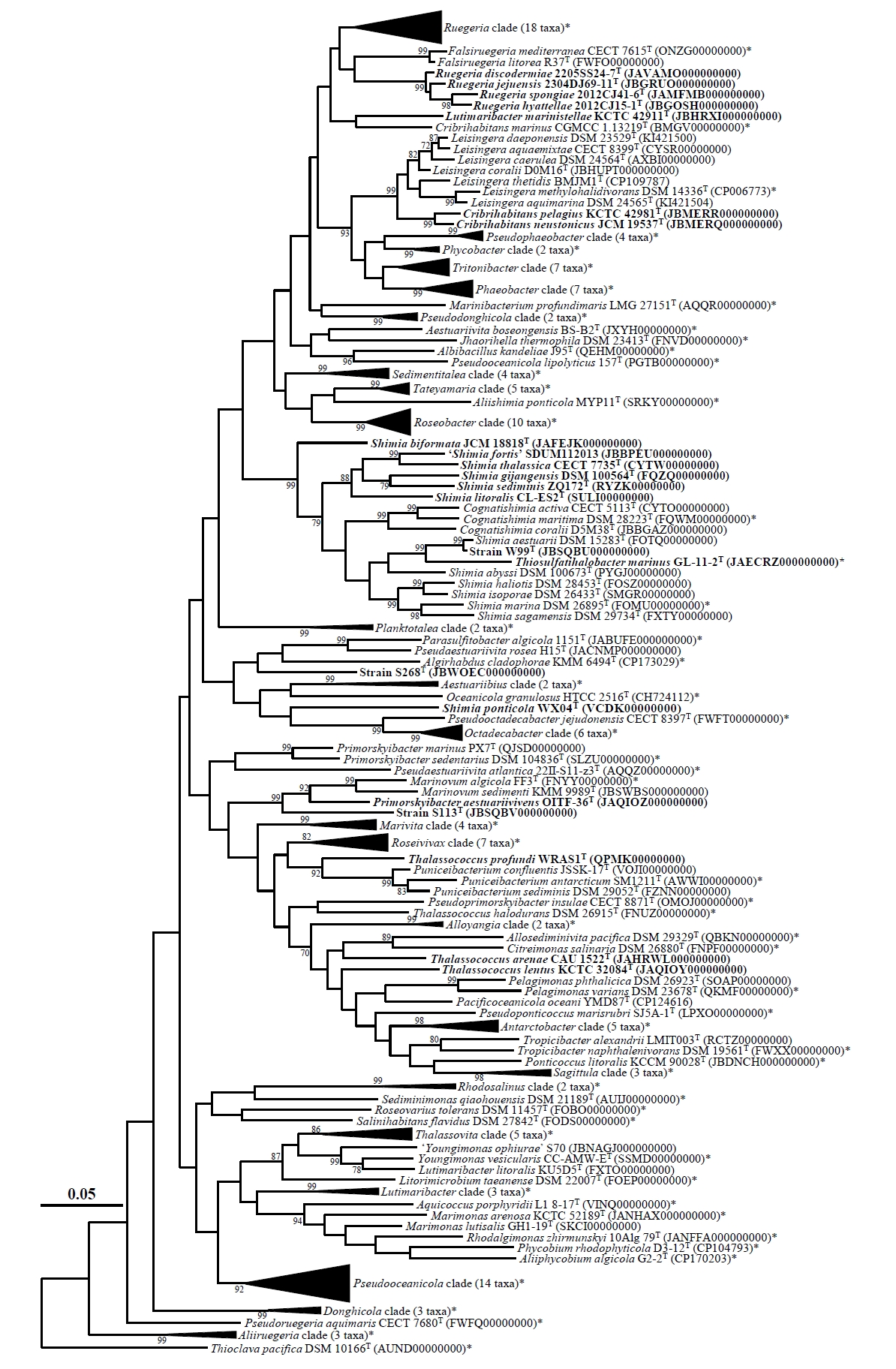
An ML phylogenomic tree based on the concatenated amino acid sequences of 120 conserved bacterial marker genes placed strain W99T within the genus Shimia, where it formed a robust clade with S. aestuarii DSM 15283T (99% bootstrap support) within the lineage containing the type species, Shimia marina (Fig. 2). This topology contrasted with the 16S rRNA gene phylogeny, in which strain W99T was separated from the type-species lineage. Notably, species currently assigned to Shimia did not form a monophyletic clade in the phylogenomic tree, supporting the need for taxonomic revision of the genus. In contrast, strains S113T and S268T each formed an independent lineage distinct from their closest relatives. Strain S113T was clearly separated from Marinovum algicola FF3T, Marinovum sedimenti KMM 9989T, and Primorskyibacter aestuariivivens OITF-36T, whereas strain S268T formed a distinct lineage separate from Parasulfitobacter algicola 1151T, Pseudaestuariivita rosea H15T, and Algirhabdus cladophorae KMM 6494T. These results support the placement of strain W99T within the genus Shimia and indicate that strains S113T and S268T represent two novel genus-level lineages within the family Roseobacteraceae.
The ANI and dDDH values between strain W99T and its closest type strain, S. aestuarii DSM 15283T, were 94.9% and 59.6%, respectively, whereas those between strain W99T and all other related type strains were ≤ 79.4% ANI and ≤ 21.8% dDDH (Table S3). Although the ANI value of 94.9% falls within the borderline range for species delineation (94–96%), the dDDH value of 59.6% is below the 70% species threshold (Riesco and Trujillo, 2024), supporting the recognition of strain W99T as a novel species of the genus Shimia. Strains S113T and S268T also exhibited low ANI and dDDH values relative to their closest type strains, with maximum values of 73.2% ANI and 24.9% dDDH for strain S113T and 71.0% ANI and 20.7% dDDH for strain S268T (Table S3). These values indicate substantial genomic divergence from currently recognized taxa. However, because universally accepted ANI and dDDH thresholds for genus delineation have not been established, these indices alone are insufficient to justify recognition of novel genera. Furthermore, clustering patterns based on ANI and dDDH as well as AAI (Fig. S2, Table S3) were not fully congruent with the phylogenomic topology (Fig. 2), indicating that genome-relatedness indices and phylogenomic analyses capture different aspects of evolutionary relationships. Together, these findings suggest that taxonomic assignments within the family Roseobacteraceae should be based on multiple complementary genomic criteria, with genome-relatedness indices interpreted in conjunction with phylogenomic evidence rather than used as standalone measures.
Several genome-based metrics have been proposed for genus delineation, among which AAI is one of the most widely used. However, no universally accepted AAI threshold for genus delineation has been established. Konstantinidis et al. (2017) reported that AAI values generally range from 65–95% among members of the same genus and 45–65% among members of the same family, whereas Luo et al. (2014) found that AAI values between members of different genera typically range from 60% to 80% and rarely exceed 85%, indicating substantial overlap among taxonomic groups. More recently, Nicholson et al. (2020) proposed that AAI values ≥ 76% support assignment to the same genus, whereas values ≤ 74% indicate different genera. Pairwise AAI analyses showed that strains S113T and S268T shared maximum AAI values of only 71.8% and 70.8%, respectively, with their closest type strains (Fig. S2). These values fall below the genus boundary proposed by Nicholson et al. (2020) and, together with their distinct phylogenomic positions, support the recognition of strains S113T and S268T as the type strains of two novel genera within the family Roseobacteraceae.
Beyond the taxonomic positions of the novel isolates, the phylogenomic analysis also revealed inconsistencies in the classification of several currently recognized taxa within the family Roseobacteraceae, particularly members of the genus Shimia. In the phylogenomic tree, species assigned to Shimia did not form a monophyletic clade but were divided into multiple lineages, with members of the genus Cognatishimia nested between Shimia clades (Fig. 2). Consistent with this topology, Cognatishimia species shared low AAI values (71.0–71.8%) with the type species, S. marina DSM 26895T, confirming their clear separation from the Shimia type-species lineage (Fig. S2A). These results indicate that the current circumscription of Shimia does not accurately reflect its phylogenomic and evolutionary relationships.
Species comprising the Shimia lineage lacking the type species, including 'Shimia fortis' SDUM112013 (Dong et al., 2026), Shimia thalassica CECT 7735T (Arahal et al., 2019), Shimia gijangensis DSM 100564T (Arahal et al., 2019), Shimia sediminis ZQ172T (Zhu et al., 2021), and Shimia litoralis CL-ES2T (Zhang et al., 2023), shared AAI values of only 72.9–73.7% with S. marina DSM 26895T, indicating substantial genomic divergence from the type-species lineage (Fig. S2A). Likewise, Shimia biformata JCM 18818T formed an independent phylogenomic lineage and shared an AAI value of only 72.5% with S. marina DSM 26895T, whereas Shimia ponticola WX04T also formed an independent lineage and shared AAI values of ≤ 68.7% with all neighboring taxa (Fig. S2A). Together with the phylogenomic evidence, these genomic characteristics support the reclassification of the type-species-lacking Shimia lineage, S. biformata, and S. ponticola into three separate novel genera distinct from the Shimia type-species lineage within the family Roseobacteraceae.
In contrast, Thiosulfatihalobacter marinus GL-11-2T, the sole validly published species of the genus Thiosulfatihalobacter (Wu et al., 2022), was nested within the Shimia sensu stricto clade containing the type species, S. marina DSM 26895T (Fig. 2). Consistent with this phylogenomic placement, Thiosulfatihalobacter marinus shared an AAI value of 75.6% with Shimia marina DSM 26895T (Fig. S2A). Although T. marinus shared an AAI value of 75.6% with S. marina DSM 26895T, which falls within the intermediate range for genus delineation (Fig. S2A), its placement within the Shimia type-species lineage provides the primary evidence for its transfer to Shimia, with the AAI value serving as complementary evidence.
Beyond the taxonomic revisions involving Shimia and Thiosulfatihalobacter, the phylogenomic analysis also revealed inconsistencies in the classification of several additional members of the family Roseobacteraceae, including twelve species with validly published names (Figs. 2 and S2). Among them, Ruegeria discodermiae 2205SS24-7T, Ruegeria jejuensis 2304DJ69-11T, Ruegeria spongiae 2012CJ41-6T, and Ruegeria hyattellae 2012CJ15-1T formed a well-supported monophyletic clade distinct from the Ruegeria sensu stricto clade containing the type species, Ruegeria atlantica CECT 4292T (Fig. 2). Instead, these four species clustered with members of the genus Falsiruegeria. They shared high AAI values among themselves (88.5–93.8%) but only 75.6–75.9% AAI with R. atlantica CECT 4292T and 75.0–75.5% with the type species of Falsiruegeria, Falsiruegeria mediterranea CECT 7615T (Fig. S2A). Although these AAI values fall within the intermediate range for genus delineation proposed by Nicholson et al. (2020), the formation of a well-supported and cohesive phylogenomic lineage distinct from the type-species lineages of both Ruegeria and Falsiruegeria provides the primary evidence for recognizing these four species as a separate genus, while their high within-lineage AAI values provide additional genomic support. Although 16S rRNA gene phylogeny has limited resolution for genus-level classification, it also supports their separation from Falsiruegeria (Figs. 1 and S1). Collectively, these findings support the reclassification of the four species into a novel genus within the family Roseobacteraceae.
Likewise, Lutimaribacter marinistellae KCTC 42911T formed an independent lineage clearly separated from the remaining members of the genus Lutimaribacter (Fig. 2). Although Lutimaribacter marinistellae was positioned closest to Cribrihabitans marinus, this relationship received low bootstrap support, and the two strains shared an AAI value of 75.3%, which falls within the intermediate range for genus delineation (Fig. S2C). Therefore, the distinct phylogenomic placement of L. marinistellae from both the Lutimaribacter type-species lineage and Cribrihabitans provides the primary evidence supporting its reclassification into a novel genus, with the AAI value serving as complementary genomic evidence. These findings indicate that Lutimaribacter marinistellae is genomically and phylogenetically distinct from both Lutimaribacter and Cribrihabitans, supporting its reclassification into a novel genus within the family Roseobacteraceae. In contrast, Cribrihabitans pelagius KCTC 42981T and Cribrihabitans neustonicus JCM 19537T formed a well-supported clade within the genus Leisingera (99% bootstrap support; Fig. 2). Consistent with this phylogenomic placement, both species shared high AAI values with Leisingera methylohalidivorans DSM 14336T, the type species of the genus Leisingera (81.8% and 81.5%, respectively; Fig. S2C). Their phylogenomic placement and AAI values therefore justify assignment of Cribrihabitans pelagius and Cribrihabitans neustonicus to Leisingera.
Furthermore, Primorskyibacter aestuariivivens OITF-36T, which was clearly separated from the remaining members of the genus Primorskyibacter, clustered within the genus Marinovum with strong bootstrap support (92%; Fig. 2). Consistent with this phylogenomic placement, it shared an AAI value of 76.8% with Marinovum algicola FF3T, the type species of the genus Marinovum, exceeding the genus boundary proposed by Nicholson et al. (2020) (Fig. S2C). Together, these findings support the transfer of Primorskyibacter aestuariivivens to the genus Marinovum. In contrast, strain S113T shared only 70.8% AAI with Marinovum algicola FF3T, well below the proposed genus boundary, and formed a distinct phylogenomic lineage separate from Marinovum. These findings indicate that strain S113T represents a novel genus rather than a member of Marinovum.
Likewise, Thalassococcus profundi WRAS1T, Thalassococcus arenae CAU 1522T, and Thalassococcus lentus KCTC 32084T each formed independent phylogenomic lineages distinct from both the Thalassococcus sensu stricto clade containing the type species and other related genera (Fig. 2). Thalassococcus profundi WRAS1T was positioned adjacent to Puniceibacterium with 92% bootstrap support but remained distinct from the lineage containing its type species, Puniceibacterium antarcticum SM1211T. Although the two strains shared an AAI value of 75.6%, which falls within the intermediate range for genus delineation (Fig. S2C), the distinct phylogenomic placement of Thalassococcus profundi provides the primary evidence for its reclassification into a novel genus within the family Roseobacteraceae, with AAI and other genome-relatedness indices serving as complementary evidence. Similarly, Thalassococcus arenae CAU 1522T and Thalassococcus lentus KCTC 32084T each formed independent phylogenomic lineages and shared AAI values of ≤ 74.3% and ≤ 75.7%, respectively, with their closest relatives (Fig. S2C). Their distinct phylogenomic placements, together with these genome-relatedness values as complementary evidence, support their recognition as representatives of two additional novel genera within the family Roseobacteraceae.
To further evaluate the consistency of the proposed taxonomic framework, pairwise genome-relatedness indices (ANI, AAI, and POCP) were compared among members of the genera affected by the proposed reclassifications before and after taxonomic revision (Fig. 3). Under the current taxonomy, intra- and intergeneric comparisons showed substantial overlap across all three indices, resulting in poorly resolved genus boundaries (Fig. 3A–3C). In contrast, the proposed reclassifications markedly reduced this overlap and produced clearer separation between intra- and intergeneric comparisons. This improvement was consistently observed in the AAI-POCP, AAI-ANI, and ANI-POCP relationships, indicating greater concordance between phylogenomic topology and genome-relatedness indices (Fig. 3D–3F). Collectively, these results provide independent quantitative support for the proposed taxonomic revisions and demonstrate that the revised classification more accurately reflects the evolutionary relationships within the family Roseobacteraceae.
Phenotypic, physiological, and biochemical characteristics
Strain W99T grew well on MA and also on NA, R2A agar, TSA, and LB agar supplemented with approximately 2% (w/v) NaCl. In contrast, strain S113T grew only on MA, whereas strain S268T grew on both MA and R2A agar. Cells of strains W99T, S113T, and S268T were Gram-stain-negative, non-motile rods measuring approximately 1.0–1.1 × 2.8–2.9 µm, 0.6–0.7 × 1.2–1.3 µm, and 0.7–0.8 × 1.4–1.5 µm, respectively (Fig. S3). None of the strains possessed flagella or exhibited gliding motility, and all were obligately aerobic.
Strains W99T, S113T, and S268T shared many phenotypic, physiological, and biochemical characteristics with their closely related species, including positive reactions for oxidase and catalase and negative reactions for indole production, glucose fermentation, arginine dihydrolase activity, hydrolysis of casein, Tween 20, and Tween 80, and assimilation of potassium gluconate and capric acid. However, they differed from their closest relatives in cell motility, nitrate reduction, urease and β-galactosidase activities, hydrolysis of esculin, gelatin, starch, and tyrosine, and assimilation of several substrates, including ᴅ-glucose, ʟ-arabinose, ᴅ-mannose, ᴅ-mannitol, N-acetyl-glucosamine, ᴅ-maltose, adipic acid, malic acid, trisodium citrate, and phenylacetic acid (Table 1).
Chemotaxonomic characteristics
All three isolates contained Q-10 as the only detectable respiratory quinone, consistent with closely related members of the family Roseobacteraceae (Nedashkovskaya et al., 2025; Park et al., 2016; Yi and Chun, 2006). The major fatty acids (> 5.0%) were 11-methyl C18:1 ω7c and summed feature 8 in strain W99T; C16:0 and summed feature 8 in strain S113T; and 11-methyl C18:1 ω7c, cyclo-C19:0 ω8c, and summed feature 8 in strain S268T. Detailed cellular fatty acid compositions of the three isolates and their closely related reference strains are presented in Table S4. The polar lipid profile of strain W99T comprised PG, PC, one unidentified phospholipid (PL), and one unidentified aminolipid (AL), consistent with those of Shimia species and Thiosulfatihalobacter marinus (Wu et al., 2022). The major polar lipids of strain S113T were PG, PC, one AL, and one unidentified lipid (L), whereas strain S268T contained PG, PC, DPG, one PL, and two ALs (Fig. S4).
Taxonomic conclusion
The combined phylogenetic, phylogenomic, genome-based, phenotypic, biochemical, and chemotaxonomic evidence demonstrates that strain W99T represents a novel species of the genus Shimia, for which the name Shimia rhodophyticola sp. nov. is proposed. Likewise, strains S113T and S268T represent two novel genus-level lineages within the family Roseobacteraceae and are proposed as Neomarinovum algicola gen. nov., sp. nov. and Phycorhabdus gracilariae gen. nov., sp. nov., respectively. In addition, comprehensive phylogenomic and genome-based relatedness analyses support the transfer of Thiosulfatihalobacter marinus to the genus Shimia. Because retention of the specific epithet marinus would create a later homonym of the pre-existing Shimia marina Choi and Cho 2006, the replacement name Shimia oceanica nom. nov. is proposed. The analyses also support the transfer of Cribrihabitans pelagius and Cribrihabitans neustonicus to the genus Leisingera as Leisingera pelagia comb. nov. and Leisingera neustonica comb. nov., respectively, and Primorskyibacter aestuariivivens to the genus Marinovum as Marinovum aestuariivivens comb. nov.
Furthermore, several phylogenetically and genomically distinct lineages are proposed as novel genera within the family Roseobacteraceae. These include Parashimia gen. nov. for five species currently assigned to Shimia (S. litoralis, 'S. fortis', S. thalassica, S. gijangensis, and S. sediminis), Pararuegeria gen. nov. for four species currently assigned to Ruegeria (R. discodermiae, R. jejuensis, R. spongiae, and R. hyattellae), Alterishimia gen. nov. for Shimia biformata, Neoshimia gen. nov. for Shimia ponticola, Paracribrihabitans gen. nov. for Lutimaribacter marinistellae, Parathalassococcus gen. nov. for Thalassococcus arenae, Neothalassococcus gen. nov. for Thalassococcus lentus, and Allothalassococcus gen. nov. for Thalassococcus profundi. A summary of the current and proposed taxonomy of the affected members of the family Roseobacteraceae is presented in Table 2.
Following the transfer of Primorskyibacter aestuariivivens, an emended description of the genus Marinovum is proposed. An emended description of the genus Shimia is also provided to reflect its revised circumscription following the transfer of Thiosulfatihalobacter marinus and the reassignment of several species to newly established genera.
Description of Shimia rhodophyticola sp. nov.
Shimia rhodophyticola (rho.do.phy.ti′co.la. N.L. neut. pl. n. Rhodophyta, the division of the red algae; L. suffix. -cola (from L. masc. or fem. n. incola), inhabitant, dweller; N.L. fem. n. rhodophyticola, inhabitant of Rhodophyta).
Colonies on MA are cream, circular, and smooth. Cells are Gram-stain-negative, obligately aerobic, and non-flagellated rods without motility. Oxidase- and catalase-positive. Growth occurs at 15–40°C (optimum, 25°C), at pH 7.0–10.0 (optimum, pH 8.0), and in the presence of 0–7.0% (w/v) NaCl (optimum, 2.0%). Nitrate is reduced to nitrite, whereas indole production and glucose fermentation are negative. Hydrolyzes tyrosine, urea, and gelatin, but not casein, Tween 20, Tween 80, esculin, or starch. Arginine dihydrolase and β-galactosidase activities are absent. Assimilates ᴅ-glucose, adipic acid, malic acid, trisodium citrate, and phenylacetic acid, but not ʟ-arabinose, ᴅ-mannose, ᴅ-mannitol, N-acetyl-glucosamine, ᴅ-maltose, potassium gluconate, or capric acid. Q-10 is the sole respiratory quinone. Major cellular fatty acids (> 5.0%) are 11-methyl C18:1 ω7c and summed feature 8. The polar lipids comprise PG, PC, an unidentified phospholipid, and an unidentified aminolipid.
The type strain is W99T (= KACC 24154T = JCM 38196T), isolated from the phycosphere of the marine red alga Schizymenia sp., collected from a coastal region in South Korea. The genome size of the strain is 4,262 kb, and its DNA G + C content is 60.5 mol%, as determined from the whole genome sequence. The GenBank accession numbers for the 16S rRNA gene and the genome sequences of strain W99T are PV993570 and JBSQBU000000000, respectively.
Description of Neomarinovum gen. nov.
Neomarinovum (Ne.o.ma.rin.o′vum. Gr. masc. adj. neos, new; N.L. neut. n. Marinovum, a bacterial genus name; N.L. neut. n. Neomarinovum, the new Marinovum).
Cells are Gram-stain-negative, aerobic, non-motile, and non-flagellated rods. Catalase- and oxidase-positive. Nitrate is not reduced. Q-10 is the major respiratory quinone. Major fatty acids (> 5.0%) are C16:0 and summed feature 8. Major polar lipids comprise PG and PC. Phylogenetically, the genus is a member of the family Roseobacteraceae within the order Rhodobacterales of the phylum Pseudomonadota. The type species is Neomarinovum algicola.
Description of Neomarinovum algicola sp. nov.
Neomarinovum algicola (al.gi′co.la. L. fem. n. alga, an alga; L. masc./fem. n. suff. -cola, inhabitant, dweller; from L. masc./fem. n. incola, dweller; N.L. masc./fem. n. algicola, an alga dweller).
In addition to the characteristics given for the genus, colonies on MA are grayish-pink, circular, and smooth. Cells are short rods (0.6–0.7 µm wide and 1.2–1.3 µm long) and do not exhibit gliding motility. Growth occurs at 20–30°C (optimum, 25°C), at pH 7.0–9.0 (optimum, pH 8.0), and in the presence of 1.0–3.0% (w/v) NaCl (optimum, 2.0%). Indole production and glucose fermentation are negative. Hydrolyzes tyrosine, but not gelatin, casein, Tween 20, Tween 80, esculin, or starch. Positive for β-galactosidase activity, but negative for urease and arginine dihydrolase activities. Does not assimilate ᴅ-glucose, ʟ-arabinose, ᴅ-mannose, ᴅ-mannitol, N-acetyl-glucosamine, ᴅ-maltose, potassium gluconate, capric acid, adipic acid, malic acid, trisodium citrate, or phenylacetic acid. Additional polar lipids include one unidentified aminolipid and one unidentified lipid.
The type strain is S113T (= KACC 23417T = JCM 38210T), isolated from the phycosphere of the marine green alga Chaetomorpha sp., collected from a coastal region in South Korea. The genome size of the strain is 4,314 kb, and its DNA G + C content is 60.0 mol%, as determined from the whole genome sequence. The GenBank accession numbers for the 16S rRNA gene and the genome sequences of strain S113T are OR234759 and JBSQBV000000000, respectively.
Description of Phycorhabdus gen. nov.
Phycorhabdus (Phy.co.rhab′dus. Gr. neut. n. phykos, algae; Gr. fem. n. rhabdos, a rod; N.L. fem. n. Phycorhabdus, a rod-shaped organism associated with algae).
Cells are Gram-stain-negative, aerobic, non-motile, and non-flagellated rods. Catalase- and oxidase-positive. Nitrate is reduced to nitrite. Q-10 is the major respiratory quinone. Major cellular fatty acids (> 5.0%) are 11-methyl C18:1 ω7c, cyclo-C19:0 ω8c, and summed feature 8. Major polar lipids comprise PG, PC, and DPG. Phylogenetically, the genus is a member of the family Roseobacteraceae within the order Rhodobacterales of the phylum Pseudomonadota. The type species is Phycorhabdus gracilariae.
Description of Phycorhabdus gracilariae sp. nov.
Phycorhabdus gracilariae (gra.ci.la′ri.ae. N.L. gen. fem. n. gracilariae, of the red alga Gracilaria).
In addition to the characteristics given for the genus, colonies on MA are pink, circular, and smooth. Cells are short rods (0.7–0.8 µm wide and 1.4–1.5 µm long) and do not exhibit gliding motility. Growth occurs at 15–40°C (optimum, 30°C), at pH 7.0–11.0 (optimum, pH 8.0), and in the presence of 1.0–7.0% (w/v) NaCl (optimum, 2.0%). Indole production and glucose fermentation are negative. Does not hydrolyze esculin, gelatin, casein, Tween 20, Tween 80, starch, or tyrosine. Positive for urease and β-galactosidase activities, but negative for arginine dihydrolase activity. Weakly assimilates ᴅ-maltose and malic acid, but does not assimilate ᴅ-glucose, ʟ-arabinose, ᴅ-mannose, ᴅ-mannitol, N-acetyl-glucosamine, potassium gluconate, capric acid, adipic acid, trisodium citrate, or phenylacetic acid. Additional polar lipids include one unidentified phospholipid and two unidentified aminolipids.
The type strain is S268T (= KACC 24156T = JCM 38211T), isolated from the phycosphere of the marine red alga Gracilaria sp., collected from a coastal region in South Korea. The genome size of the strain is 3,864 kb, and its DNA G + C content is 56.1 mol%, as determined from the whole genome sequence. The GenBank accession numbers for the 16S rRNA gene and the genome sequences of strain S268T are PV993569 and JBWOEC000000000, respectively.
Description of Pararuegeria gen. nov.
Pararuegeria (Pa.ra.rue.ge′ri.a. Gr. prep. para, beside, alongside of, near; N.L. fem. n. Ruegeria, a bacterial generic name; N.L. fem. n. Pararuegeria, a genus alongside, or closely related to, the genus Ruegeria).
Cells are Gram-stain-negative, aerobic, and ovoid to short rod-shaped. Motility by flagella is variable. Oxidase and catalase are positive, whereas nitrate reduction is variable. The genomic DNA G + C content ranges from 59.0 to 60.8 mol%. The predominant respiratory quinone is Q-10. The major cellular fatty acids (> 5.0%) are C12:0 3-OH, C16:0 2-OH, and summed feature 8. The major polar lipids are PC and PG, whereas PE and DPG are variably present among species. Phylogenetically, the genus belongs to the family Roseobacteraceae within the order Rhodobacterales of the phylum Pseudomonadota. The type species is Pararuegeria spongiae.
Description of Pararuegeria discodermiae comb. nov.
Pararuegeria discodermiae (dis.co.der′mi.ae. N.L. gen. n. discodermiae, of the sponge genus Discodermia).
Basonym: Ruegeria discodermiae Moon et al. 2026 (Oren and Göker, 2026).
The description of Pararuegeria discodermiae is identical to that proposed for Ruegeria discodermiae (Moon et al., 2025). The type strain is 2205SS24-7T (= KACC 23237T = LMG 33064T).
Description of Pararuegeria jejuensis comb. nov.
Pararuegeria jejuensis (je.ju.en′sis. N.L. fem. adj. jejuensis, Jeju island, Republic of Korea, from where the type strain was isolated).
Basonym: Ruegeria jejuensis Moon et al. 2026 (Oren and Göker, 2026).
The description of Pararuegeria jejuensis is identical to that proposed for Ruegeria jejuensis (Moon et al., 2025). The type strain is 2304DJ69-11T (= KACC 23805T = MCCC 1K09534T).
Description of Pararuegeria spongiae comb. nov.
Pararuegeria spongiae (spon′gi.ae. L. gen. n. spongiae, of a sponge, source of the type strain).
Basonym: Ruegeria spongiae Han and Park 2023.
The description of Pararuegeria spongiae is identical to that proposed for Ruegeria spongiae (Han and Park, 2023). The type strain is 2012CJ41-6T (= KACC 22645T = LMG 32585T).
Description of Pararuegeria hyattellae comb. nov.
Pararuegeria hyattellae (hy.at.tel′lae. N.L. gen. n. hyattellae, of the sponge genus Hyattella).
Basonym: Ruegeria hyattellae Moon et al. 2026 (Oren and Göker, 2026).
The description of Pararuegeria hyattellae is identical to that proposed for Ruegeria hyattellae (Moon et al., 2025). The type strain is 2012CJ15-1T (= KACC 23842T = MCCC 1K09526T).
Description of Paracribrihabitans gen. nov.
Paracribrihabitans (Pa.ra.cri.bri.ha′bi.tans. Gr. prep. para, beside, alongside of, near; N.L. masc. n. Cribrihabitans, a bacterial generic name; N.L. masc. n. Paracribrihabitans, a genus alongside, or closely related to, the genus Cribrihabitans).
Cells are Gram-stain-negative, aerobic, and motile rods with flagella. Catalase- and oxidase-positive. Nitrate is not reduced. Q-10 is the major respiratory quinone. Major fatty acids (> 5.0%) are 11-methyl C18:1 ω7c and summed feature 8. Major polar lipids comprise PG, DPG, and PE. Phylogenetically, the genus is a member of the family Roseobacteraceae within the order Rhodobacterales of the phylum Pseudomonadota. The type species is Paracribrihabitans marinistellae.
Description of Paracribrihabitans marinistellae comb. nov.
Paracribrihabitans marinistellae (ma.ri.ni.stel′lae. L. masc. adj. marinus, from the sea; L. fem. n. stella, star; N.L. gen. n. marinistellae, of a sea star).
Basonym: Lutimaribacter marinistellae Zhang et al. 2016.
The description of Paracribrihabitans marinistellae is identical to that proposed for Lutimaribacter marinistellae (Zhang et al., 2016). The type strain is SF-12T (= KCTC 42911T = MCCC 1K01154T).
Description of Leisingera pelagia comb. nov.
Leisingera pelagia (pe.la′gi.a. L. fem. adj. pelagia, belonging to the sea).
Basonym: Cribrihabitans pelagius Yoon et al. 2016.
The description of Leisingera pelagia is identical to that proposed for Cribrihabitans pelagius (Yoon et al., 2016). The type strain is KMU-32T (= KCTC 42981T = NBRC 111834T).
Description of Leisingera neustonica comb. nov.
Leisingera neustonica (neus.to′ni.ca. N.L. fem. adj. neustonica, pertaining to and living in the neuston).
Basonym: Cribrihabitans neustonicus Hameed et al. 2015.
The description of Leisingera neustonica is identical to that proposed for Cribrihabitans neustonicus (Hameed et al., 2014). The type strain is CC-AMHB-3T (= BCRC 80695T = JCM 19537T).
Description of Alterishimia gen. nov.
Alterishimia (Al.te.ri.shi′mi.a. L. masc. adj. alter, another, other; N.L. fem. n. Shimia, a bacterial genus name; N.L. fem. n. Alterishimia, another Shimia).
Cells are Gram-stain-negative, strictly aerobic, and non-flagellated rods. Catalase- and oxidase-positive. Nitrate is reduced to nitrite. Q-10 is the major respiratory quinone. The major cellular fatty acids (> 5.0%) are C16:0, 11-methyl C18:1 ω7c, and summed feature 8. The major polar lipids comprise DPG, PG, and PC. Phylogenetically, the genus is a member of the family Roseobacteraceae within the order Rhodobacterales of the phylum Pseudomonadota. The type species is Alterishimia biformata.
Description of Alterishimia biformata comb. nov.
Alterishimia biformata (bi.for.ma′ta. L. fem. adj. biformata, double-formed, two-shaped, pertaining to the different cell morphology during different growth phases).
Basonym: Shimia biformata Hameed et al. 2013.
The description of Alterishimia biformata is identical to that proposed for Shimia biformata (Hameed et al., 2013). The type strain is CC-AMW-CT (= BCRC 80548T = JCM 18818T).
Description of Parashimia gen. nov.
Parashimia (Pa.ra.shi′mi.a. Gr. prep. para, beside, alongside of, near; N.L. fem. n. Shimia, a bacterial generic name; N.L. fem. n. Parashimia, a genus alongside, or closely related to, the genus Shimia).
Cells are Gram-stain-negative, aerobic, and rod- or ovoid-shaped. Motility is variable. Catalase activity and nitrate reduction are variable. The genomic DNA G + C content ranges from 47.0 to 59.8 mol%. The major respiratory quinone is Q-10. The major cellular fatty acids (> 5.0%) commonly include summed feature 8. The major polar lipids commonly comprise PG, whereas PC, PE, DPG, and phosphatidyl-N,N-dimethylethanolamine are variably present depending on the strain. Phylogenetically, the genus is a member of the family Roseobacteraceae within the order Rhodobacterales of the phylum Pseudomonadota. The type species is Parashimia litoralis.
Description of Parashimia litoralis comb. nov.
Parashimia litoralis (li.to.ra′lis. L. fem. adj. litoralis, of the shore).
Basonym: Shimia litoralis (Kim et al. 2008) Zhang et al. 2023.
The description of Parashimia litoralis is identical to that proposed for Pelagicola litoralis (Kim et al., 2008). The type strain is CL-ES2T (= DSM 18290T = KCCM 42274T).
Description of Parashimia fortis sp. nov.
Parashimia fortis (for′tis. L. fem. adj. fortis, strong, referring to the fact that the strain is still viable after six months of laboratory cultivation).
Colonies on MA are beige, circular, convex, and smooth. Cells are Gram-stain-negative, aerobic, non-motile, and ovoid. Growth occurs at 20–37°C (optimum, 28–35°C) and pH 6.0–9.0 (optimum, pH 7.0) and in the presence of 1.0–7.0% (w/v) NaCl (optimum, 3.0–4.0%). Catalase activity is negative. Positive for alkaline phosphatase, esterase (C4), esterase lipase (C8), leucine arylamidase, and naphthol-AS-BI-phosphohydrolase activities, and weakly positive for valine arylamidase activity. Hydrolyzes starch, alginate, and casein, but does not hydrolyze CM-cellulose or Tweens 20, 40, 60, or 80. Acids are produced from ᴅ-ribose, fructose, ʟ-sorbose, esculin, ᴅ-tagatose, and potassium 5-ketogluconate, and weakly from ᴅ-arabinose, ʟ-arabinose, ᴅ-xylose, ʟ-xylose, ᴅ-turanose, ᴅ-lyxose, and potassium 2-ketogluconate. Utilizes glucuronamide, ʟ-malic acid, bromo-succinic acid, acetic acid, acetoacetic acid, and α-hydroxybutyric acid as sole carbon sources, and weakly utilizes ᴅ-glucuronic acid. Q-10 is the sole respiratory quinone. Major cellular fatty acids (> 5.0%) are C16:0, C17:0, 11-methyl C18:1 ω7c, and summed feature 8 (C18:1 ω7c and/or C18:1 ω6c). The polar lipids comprise PG, PME, DPG, and PE.
The type strain is SDUM112013T (= KCTC 8307T = MCCC 1H01481T), which was isolated from intertidal sediments of Xiaoshi Island, China. The genome size of the strain is 3,823 kb, and its DNA G + C content is 59.8 mol%, as determined from the whole genome sequence. The GenBank accession numbers for the 16S rRNA gene and the genome sequences of strain SDUM112013T are PP892595 and JBBPEU000000000, respectively.
Description of Parashimia thalassica comb. nov.
Parashimia thalassica (tha.las′si.ca. Gr. fem. n. thalassa, the sea; L. fem. adj. suff. -ica, suffix used with the sense of pertaining to; L. fem. adj. thalassica, pertaining to the sea, of the sea).
Basonym: Shimia thalassica Arahal et al. 2019.
The description of Parashimia thalassica is identical to that proposed for Shimia thalassica (Arahal et al., 2019). The type strain is R47T (= CECT 7735T = LMG 29909T).
Description of Parashimia gijangensis comb. nov.
Parashimia gijangensis (gi.jang.en′sis. N.L. fem. adj. gijangensis, pertaining to Gijang, the location of the National Fisheries Research and Development Institute, where the type strain was characterized).
Basonym: Shimia gijangensis (Kim et al. 2014) Arahal et al. 2019.
The description of Parashimia gijangensis is identical to that proposed for Pseudopelagicola gijangensis (Kim et al., 2014). The type strain is YSS-7T (= KCTC 42049T = CECT 8540T).
Description of Parashimia sediminis comb. nov.
Parashimia sediminis (se.di′mi.nis. L. gen. n. sediminis, of sediment).
Basonym: Shimia sediminis Zhu et al. 2021.
The description of Parashimia sediminis is identical to that proposed for Shimia sediminis (Zhu et al., 2021). The type strain is ZQ172T (= KCTC 62578T = MCCC 1K03531T).
Description of Shimia oceanica nom. nov.
Shimia oceanica (o.ce.a′ni.ca. N.L. fem. adj. oceanica, of or pertaining to the ocean).
Basonym: Thiosulfatihalobacter marinus Wu et al. 2022.
The description of Shimia oceanica is identical to that proposed for Thiosulfatihalobacter marinus (Wu et al., 2022). The type strain is GL-11-2T (= KCTC 82723T = MCCC M20691T).
Emended description of the genus Shimia Choi and Cho 2006
Emended description of the genus Shimia is as given previously by Choi and Cho (2006), with the following amendments. The major fatty acid is typically summed feature 8. Depending on the species, C16:0, 11-methyl C18:1 ω7c, or cyclo-C19:0 ω8c may occur as additional major fatty acids.
Description of Neoshimia gen. nov.
Neoshimia (Ne.o.shi′mi.a. Gr. masc. adj. neos, new; N.L. fem. n. Shimia, a bacterial genus name; N.L. fem. n. Neoshimia, the new Shimia).
Cells are Gram-stain-negative, aerobic, non-motile, and non-flagellated rods. Catalase- and oxidase-negative. Nitrate is not reduced. Q-10 is the major respiratory quinone. Major fatty acids (> 5.0%) are C18:0, 11-methyl C18:1 ω7c, C10:0 3-OH, and summed feature 8. Major polar lipids comprise PC, PE, and PG. Phylogenetically, the genus is a member of the family Roseobacteraceae within the order Rhodobacterales of the phylum Pseudomonadota. The type species is Neoshimia ponticola.
Description of Neoshimia ponticola comb. nov.
Neoshimia ponticola (pon.ti′co.la. L. masc. n. pontus, the sea; L. suff. -cola (from L. masc. or fem. n. incola) inhabitant, dweller; N.L. fem. n. ponticola, a dweller of sea).
Basonym: Shimia ponticola Chen et al. 2023.
The description of Neoshimia ponticola is identical to that proposed for Shimia ponticola (Chen et al., 2023). The type strain is WX04T (= KCTC 62628T = MCCC 1K02295T).
Description of Marinovum aestuariivivens comb. nov.
Marinovum aestuariivivens (aes.tu.a.ri.i.vi′vens. L. neut. n. aestuarium (gen. aestuarii), tidal flat; L. pres. part. vivens, living; N.L. masc. part. adj. aestuariivivens, living in a tidal flat).
Basonym: Primorskyibacter aestuariivivens Park et al. 2016.
The description of Marinovum aestuariivivens is identical to that proposed for Primorskyibacter aestuariivivens (Park et al., 2016). The type strain is OITF-36T (= KCTC 52455T = NBRC 112419T).
Emended description of the genus Marinovum Martens et al. 2006S
The description of the genus is as given previously by Lafay et al. (1995) and Martens et al. (2006), with the following amendments. The major fatty acid is C18:1 ω7c. Major polar lipids are typically PG, PE, and PC; depending on the species, additional polar lipids such as DPG or phosphatidylmonomethylethanolamine may also be present.
Description of Allothalassococcus gen. nov.
Allothalassococcus (Al.lo.tha.las.so.coc′cus. Gr. adj. allos, other; N.L. masc. n. Thalassococcus, a bacterial genus name; N.L. masc. n. Allothalassococcus, another Thalassococcus).
Cells are Gram-stain-negative, aerobic, non-motile, and rod- or ovoid-shaped. Catalase- and oxidase-positive. Nitrate is not reduced. Q-10 is the major respiratory quinone. Major fatty acids (> 5.0%) are C12:1 3-OH, C16:0, and summed feature 8. Major polar lipids are PC, PG, and PE. Phylogenetically, the genus is a member of the family Roseobacteraceae within the order Rhodobacterales of the phylum Pseudomonadota. The type species is Allothalassococcus profundi.
Description of Allothalassococcus profundi comb. nov.
Allothalassococcus profundi (pro.fun′di. L. gen. n. profundi, of/ from the depths of the sea).
Basonym: Thalassococcus profundi Yu et al. 2019.
The description of Allothalassococcus profundi is identical to that proposed for Thalassococcus profundi (Yu et al., 2019). The type strain is WRAS1T (= KCTC 52696T = MCCC 1K03253T).
Description of Parathalassococcus gen. nov.
Parathalassococcus (Pa.ra.tha.las.so.coc′cus. Gr. prep. para, beside; N.L. masc. n. Thalassococcus, a bacterial genus name; N.L. masc. n. Parathalassococcus, a genus beside Thalassococcus).
Cells are Gram-stain-negative, aerobic, and non-motile rods. Catalase- and oxidase-positive. Nitrate is reduced. Q-10 is the major respiratory quinone. Major fatty acids (> 5.0%) are 11-methyl C18:1 ω7c, C18:0, C17:0, and summed feature 8. Major polar lipids are PC, PE, and PG. Phylogenetically, the genus is a member of the family Roseobacteraceae within the order Rhodobacterales of the phylum Pseudomonadota. The type species is Parathalassococcus arenae.
Description of Parathalassococcus arenae comb. nov.
Parathalassococcus arenae (a.re′nae. L. gen. n. arenae, of sand).
Basonym: Thalassococcus arenae Weerawongwiwat et al. 2024 (Oren and Göker, 2024).
The description of Parathalassococcus arenae is identical to that proposed for Thalassococcus arenae (Weerawongwiwat et al., 2022). The type strain is CAU 1522T (= KCTC 72545T = MCCC 1K04064T).
Description of Neothalassococcus gen. nov.
Neothalassococcus (Ne.o.tha.las.so.coc′cus. Gr. masc. adj. neos, new; N.L. masc. n. Thalassococcus, a bacterial genus name; N.L. masc. n. Neothalassococcus, a new Thalassococcus).
Cells are Gram-stain-negative, aerobic, non-motile, and rod- or ovoid-shaped. Catalase-positive and oxidase-negative. Nitrate is not reduced. Q-10 is the major respiratory quinone. Major fatty acids (> 5.0%) are 11-methyl C18:1 ω7c, C18:1 ω7c, and C18:0. Major polar lipids are PC and PG. Phylogenetically, the genus is a member of the family Roseobacteraceae within the order Rhodobacterales of the phylum Pseudomonadota. The type species is Neothalassococcus lentus.
Description of Neothalassococcus lentus comb. nov.
Neothalassococcus lentus (len′tus. L. masc. adj. lentus, slow, delayed, referring to the slow growth of the microorganism).
Basonym: Thalassococcus lentus Park et al. 2013.
The description of Neothalassococcus lentus is identical to that proposed for Thalassococcus lentus (Park et al., 2013). The type strain is YCS-24T (= CCUG 62791T = KCTC 32084T).
Acknowledgments
This work was supported by the Marine Biotics Project (20210469) funded by the Ministry of Oceans and Fisheries, Republic of Korea, and the National Institute of Biological Resources (202602204), funded by the Ministry of Climate, Energy, Environment, Republic of Korea. We thank Dr. Aharon Oren (The Hebrew University of Jerusalem, Israel) for his valuable advice on the etymology.
Conflict of Interest
The authors declare no competing financial conflicts of interest.
Data Availability
The GenBank accession numbers for the 16S rRNA gene sequences of strains W99T, S113T, and S268T are PV993570, OR234759, and PV993569, respectively. The genome sequences for strains W99T, S113T, and S268T are available under the accession numbers JBSQBU000000000, JBSQBV000000000, and JBWOEC000000000, respectively.
Supplementary Information
The online version contains supplementary material available at https://doi.org/10.71150/jm.2607010
Table S1.
Potential ecological distributions of 16S rRNA gene sequences closely related to strains W99T, S113T, and S268T, assessed using the Integrated Microbial Next-Generation Sequencing (IMNGS) platform at a 99.0% sequence similarity threshold. “Matched no. (Prevalence)” represents the number (percentage) of datasets containing 16S rRNA gene sequences showing ≥ 99.0% sequence similarity to the respective query sequences of strain W99T, S113T, and S268T, and “ARA” denotes the average relative abundance of the matched sequences within the datasets. Prevalence values below 0.5% for all three query sequences are not shown. –, not detected.
jm-2607010-Supplementary-Table-S1.pdf
Table S3.
Average nucleotide identity (ANI; lower-left triangle) and digital DNA–DNA hybridization (dDDH; upper-right triangle) values among strains W99T, S113T, and S268T and their closely related type strains
jm-2607010-Supplementary-Table-S3.pdf
Fig. S1.
Maximum-likelihood (ML; A) and maximum-parsimony (MP; B) trees based on 16S rRNA gene sequences showing the phylogenetic positions of strains W99T, S113T, and S268T (shown in bold) within the family Roseobacteraceae. Bootstrap values > 70%, calculated from 1,000 replicates, are shown at the nodes. Thioclava pacifica TL 2T (KX618934) was used as the outgroup. Type species are indicated by asterisks, and black triangles represent collapsed clades containing the corresponding type species. Scale bars represent nucleotide substitutions per site in the ML tree and the number of character changes across the full 16S rRNA gene sequence in the MP tree.
jm-2607010-Supplementary-Fig-S1.pdf
Fig. S2.
Average amino acid identity (AAI) values among strains W99T and S268T and their closely related taxa (A), among strain S113T and its closely related taxa (B), and among taxa of other clades in the phylogenomic tree (C). Dendrograms were generated by hierarchical clustering of AAI values using the ComplexHeatmap package (version 2.14.0) in R.
jm-2607010-Supplementary-Fig-S2.pdf
Fig. S3.
Transmission electron micrograph showing the general morphology of negatively stained cells of strains W99T (A), S113T (B), and S268T (C). Cells were grown on marine agar at their respective optimum temperatures for 3 days and negatively stained with UranyLess.
jm-2607010-Supplementary-Fig-S3.pdf
Fig. S4.
Two-dimensional thin-layer chromatograms (TLC) showing the polar lipid profile of strains W99T, S113T, and S268T. Solvent systems: (I) chloroform-methanol-water (65:25:4, v/v/v) and (II) chloroform-acetic acid-methanol-water (80:15:12:4, v/v/v/v). The TLC plates were sprayed with 10% ethanolic molybdophosphoric acid (A), ninhydrin (B), Dittmer-Lester (C), and α-naphthol/sulfuric acid (D) reagents for the detection of total polar lipids, aminolipids, phospholipids, and glycolipids, respectively. PG, phosphatidylglycerol; PC, phosphatidylcholine; DPG, diphosphatidylglycerol; PL, unidentified phospholipid; AL, unidentified aminolipid; L, unidentified lipid.
jm-2607010-Supplementary-Fig-S4.pdf
Fig. 1.Neighbor-joining tree based on 16S rRNA gene sequences showing the phylogenetic positions of strains W99T, S113T, and S268T (shown in bold) within the family Roseobacteraceae. Bootstrap values > 70%, calculated from 1,000 replicates, are shown at the nodes. Thioclava pacifica TL 2T (KX618934) was used as the outgroup. Type species are indicated by asterisks, and black triangles represent collapsed clades containing the corresponding type species. The scale bar indicates 0.01 nucleotide substitutions per site.

Fig. 2.Maximum-likelihood phylogenomic tree showing the positions of strains W99T, S113T, and S268T (shown in bold) within the family Roseobacteraceae, based on the concatenated amino acid sequences of 120 bacterial marker genes identified by GTDB-Tk. Bootstrap values > 70%, calculated from 1,000 replicates, are shown at the nodes. Thioclava pacifica DSM 10166T (AUND00000000) was used as the outgroup. Type species are indicated by asterisks, and black triangles represent collapsed clades containing the corresponding type species. Taxa that may warrant taxonomic reclassification based on the phylogenomic topology are also shown in bold. The scale bar indicates 0.05 amino acid substitutions per site.

Fig. 3.Pairwise comparisons of genome-relatedness indices among members of genera affected by the proposed taxonomic revisions within the family Roseobacteraceae, based on the current and proposed classifications. Relationships between average amino acid identity (AAI) and percentage of conserved proteins (POCP) (A, D), AAI and average nucleotide identity (ANI) (B, E), and ANI and POCP (C, F) are shown for the current taxonomy (A–C) and the proposed taxonomy (D–F). Red and blue symbols represent intergeneric and intrageneric comparisons, respectively.

Table 1.
Differential phenotypic characteristics of strains W99T, S113T, and S268T and closely related taxa within the family Roseobacteraceae
Taxa: 1, strain W99ᵀ (this study); 2, strain S113ᵀ (this study); 3, strain S268ᵀ (this study); 4, Shimia aestuarii KCTC 12049T (Wirth and Whitman, 2018; Yi and Chun, 2006); 5, Thiosulfatihalobacter marinus KCTC 82723T (Wu et al., 2022); 6, Marinovum algicola JCM 21266T (Lafay et al., 1995; Martens et al., 2006); 7, Primorskyibacter aestuariivivens KCTC 52455T (Park et al., 2016). All strains are positive for oxidase and catalase activities*. All strains are negative for the following characteristics: indole production*, glucose fermentation*, hydrolysis* of casein, Tween 20, and Tween 80, arginine dihydrolase activity*, and assimilation* of potassium gluconate and capric acid. +, positive; –, negative; w, weakly positive; ND, no data.
|
Characteristic |
1 |
2 |
3 |
4 |
5 |
6 |
7 |
|
Isolation source |
Marine alga |
Marine alga |
Marine alga |
Tidal sediment |
Seawater |
Dinoflagellate |
Tidal flat |
|
Colony color |
Cream |
Grayish pink |
Pink |
Cream |
Cream |
White |
Greyish yellow |
|
Cell morphology |
Short rod |
Short rod |
Short rod |
Ovoid |
Rod |
Ovoid rod |
Ovoid or rod |
|
Cell motility |
− |
− |
− |
− |
− |
+ |
+ |
|
Growth condition: |
|
|
|
|
|
|
|
|
Temperature (°C, optimum) |
15–40 (25) |
20–30 (25) |
15–40 (30) |
15–35 (35) |
4–40 (30) |
10–37 (25–30) |
10–40 (30) |
|
pH (optimum) |
7.0–10.0 (8.0) |
7.0–9.0 (8.0) |
7.0–11.0 (8.0) |
6.0–11.0 (7.0) |
6.0–8.0 (6.5–7.0) |
6.0–9.0 (7.5) |
6.0–8.0 (7.0–8.0) |
|
NaCl (%, optimum) |
0–7.0 (2.0) |
1.0–3.0 (2.0) |
1.0–7.0 (2.0) |
1.0–7.0 (2.0) |
0.5–7.0 (2.0) |
0.6–11.7 (ND) |
0–9.0 (2.0–3.0) |
|
Reduction of nitrate |
+ |
− |
+ |
+ |
− |
− |
+ |
|
Hydrolysis* of: |
|
|
|
|
|
|
|
|
Esculin |
− |
− |
− |
− |
− |
+ |
+ |
|
Gelatin |
+ |
− |
− |
− |
− |
− |
− |
|
Starch |
− |
− |
− |
− |
− |
+ |
+ |
|
Tyrosine |
+ |
+ |
− |
+ |
w
|
+ |
+ |
|
Activity* of: |
|
|
|
|
|
|
|
|
Urease |
+ |
− |
+ |
− |
− |
− |
− |
|
β-Galactosidase |
− |
+ |
+ |
− |
− |
+ |
− |
|
Assimilation* of: |
|
|
|
|
|
|
|
|
ᴅ-Glucose |
+ |
− |
− |
w
|
− |
+ |
− |
|
ʟ-Arabinose |
− |
− |
− |
− |
− |
+ |
− |
|
ᴅ-Mannose |
− |
− |
− |
− |
− |
+ |
− |
|
ᴅ-Mannitol |
− |
− |
− |
− |
− |
+
|
− |
|
N-Acetyl-glucosamine |
− |
− |
− |
− |
w
|
+
|
− |
|
ᴅ-Maltose |
− |
− |
w
|
− |
− |
w
|
− |
|
Adipic acid |
w
|
− |
− |
+ |
+ |
w
|
w
|
|
Malic acid |
+ |
− |
w
|
+ |
+ |
+ |
+ |
|
Trisodium citrate |
+ |
− |
− |
+ |
w
|
+ |
w
|
|
Phenylacetic acid |
+ |
− |
− |
+ |
w
|
w
|
− |
|
Major polar lipids†
|
PC, PG |
PC, PG |
PC, PG, DPG |
ND |
PC, PG, PE, DPG |
ND |
PC, PG, PE, PME |
Table 2.Original, revised, and proposed taxonomy of species within the family Roseobacteraceae. The proposed taxonomic changes are based on the phylogenomic and genome-based relatedness analyses performed in this study. –, no taxonomic change.
|
Original taxonomy |
Revised taxonomy |
Proposed taxonomy |
|
Ruegeria discodermiae Moon et al. 2026 |
– |
Pararuegeria discodermiae comb. nov. |
|
Ruegeria jejuensis Moon et al. 2026 |
– |
Pararuegeria jejuensis comb. nov. |
|
Ruegeria spongiae Han and Park 2023 |
– |
Pararuegeria spongiae gen. nov., comb. nov. |
|
Ruegeria hyattellae Moon et al. 2026 |
– |
Pararuegeria hyattellae comb. nov. |
|
Lutimaribacter marinistellae Zhang et al. 2016 |
– |
Paracribrihabitans marinistellae gen. nov., comb. nov. |
|
Cribrihabitans pelagius Yoon et al. 2016 |
– |
Leisingera pelagia comb. nov. |
|
Cribrihabitans neustonicus Hameed et al. 2014 |
– |
Leisingera neustonica comb. nov. |
|
Shimia biformata Hameed et al. 2013 |
– |
Alterishimia biformata gen. nov., comb. nov. |
|
'Shimia fortis' Dong et al. 2026 |
– |
Parashimia fortis sp. nov. |
|
Shimia thalassica Arahal et al. 2019 |
– |
Parashimia thalassica comb. nov. |
|
Pseudopelagicola gijangensis Kim et al. 2014 |
Shimia gijangensis Arahal et al. 2019 |
Parashimia gijangensis comb. nov. |
|
Shimia sediminis Zhu et al. 2021 |
– |
Parashimia sediminis comb. nov. |
|
Pelagicola litoralis Kim et al. 2008 |
Shimia litoralis Zhang et al. 2023 |
Parashimia litoralis gen. nov., comb. nov. |
|
Thiosulfatihalobacter marinus Wu et al. 2022 |
– |
Shimia oceanica nom. nov. |
|
Shimia ponticola Chen et al. 2023 |
– |
Neoshimia ponticola gen. nov., comb. nov. |
|
Primorskyibacter aestuariivivens Park et al. 2016 |
– |
Marinovum aestuariivivens comb. nov. |
|
Thalassococcus profundi Yu et al. 2019 |
– |
Allothalassococcus profundi gen. nov., comb. nov. |
|
Thalassococcus arenae Weerawongwiwat et al. 2024 |
– |
Parathalassococcus arenae gen. nov., comb. nov. |
|
Thalassococcus lentus Park et al. 2013 |
– |
Neothalassococcus lentus gen. nov., comb. nov. |
References
- Arahal DR, La Mura A, Lucena T, Rodrigo-Torres L, Aznar R. 2019. Shimia thalassica sp. nov., and reclassification of Pseudopelagicola gijangensis as Shimia gijangensis comb. nov., and Thalassobius activus as Cognatishimia activa comb. nov. Int J Syst Evol Microbiol. 69: 3405–3413. ArticlePubMed
- Bayburt H, Choi BJ, Kim JM, Baek JH, Jeon CO. 2024. Psychrosphaera algicola sp. nov. and Paraglaciecola algarum sp. nov., and reclassification of Pseudoalteromonas elyakovii, Pseudoalteromonas flavipulchra, and Pseudoalteromonas profundi as later heterotypic synonyms of P. distincta, P. maricaloris, and P. gelatinilytica. Int J Syst Evol Microbiol. 74: 006491.ArticlePubMedPMC
- Bayburt H, Kim JM, Choi BJ, Lee JK, Jeon CO. 2026. Ningiella algicola sp. nov. and Marinicella algicola sp. nov., proposal of Paralteromonas gen. nov. and Neoalteromonas gen. nov. with reclassification of Alteromonas species, and reclassification of Methylophaga aminisulfidivorans as a later heterotypic synonym of Methylophaga thalassica. Syst Appl Microbiol. 49: 126685.ArticlePubMed
- Benmeradi N, Payre B, Goodman SL. 2015. Easier and safer biological staining: High contrast UranyLess staining of TEM grids using mPrep/g capsules. Microsc Microanal. 21: 721–722. Article
- Bernardet JF, Nakagawa Y, Holmes B. 2002. Proposed minimal standards for describing new taxa of the family Flavobacteriaceae and emended description of the family. Int J Syst Evol Microbiol. 52: 1049–1070. ArticlePubMed
- Chalita M, Kim YO, Park S, Oh HS, Cho JH, et al. 2024. EzBioCloud: A genome-driven database and platform for microbiome identification and discovery. Int J Syst Evol Microbiol. 74: 006421.ArticlePubMedPMC
- Chaumeil PA, Mussig AJ, Hugenholtz P, Parks DH. 2022. GTDB-Tk v2: Memory friendly classification with the genome taxonomy database. Bioinformatics. 38: 5315–5316. ArticlePubMedPMCPDF
- Chen Q, He B, Lin TH, Jiao N, Zheng Q. 2023. Shimia ponticola sp. nov., an aerobic anoxygenic photoheterotrophic bacterium, isolated from surface seawater in the South China Sea. Int J Syst Evol Microbiol. 73: 005859.Article
- Chen MH, Sheu SY, Chen CA, Wang JT, Chen WM. 2011. Shimia isoporae sp. nov., isolated from the reef-building coral Isopora palifera. Int J Syst Evol Microbiol. 61: 823–827. ArticlePubMed
- Chklovski A, Parks DH, Woodcroft BJ, Tyson GW. 2023. CheckM2: A rapid, scalable and accurate tool for assessing microbial genome quality using machine learning. Nat Methods. 20: 1203–1212. ArticlePubMedPDF
- Choi DH, Cho BC. 2006. Shimia marina gen. nov., sp. nov., a novel bacterium of the Roseobacter clade isolated from biofilm in a coastal fish farm. Int J Syst Evol Microbiol. 56: 1869–1873. ArticlePubMed
- Coe LS, Fei C, Weston J, Amin SA. 2023. Phycobacter azelaicus gen. nov. sp. nov., a diatom symbiont isolated from the phycosphere of Asterionellopsis glacialis. Int J Syst Evol Microbiol. 73: 006104.Article
- Dong XY, Lin PR, Tan XY, Li JQ, Du ZJ, et al. 2026. Shimia fortis sp. nov., isolated from marine sediment. Curr Microbiol. 83: 140.ArticlePubMedPDF
- Hameed A, Shahina M, Lin SY, Lai WA, Hsu YH, et al. 2013. Shimia biformata sp. nov., isolated from surface seawater, and emended description of the genus Shimia Choi and Cho 2006. Int J Syst Evol Microbiol. 63: 4533–4539. ArticlePubMed
- Hameed A, Shahina M, Lin SY, Lai WA, Liu YC, et al. 2014. Cribrihabitans neustonicus sp. nov., isolated from coastal surface seawater, and emended description of the genus Cribrihabitans Chen et al. 2014. Int J Syst Evol Microbiol. 64: 3897–3903. ArticlePubMed
- Han SM, Park JS. 2023. Ruegeria spongiae sp. nov., isolated from Callyspongia elongata. Int J Syst Evol Microbiol. 73: 006001.Article
- Hwang CY, Cho BC. 2008. Ponticoccus litoralis gen. nov., sp. nov., a marine bacterium in the family Rhodobacteraceae. Int J Syst Evol Microbiol. 58: 1332–1338. ArticlePubMed
- Hyun DW, Kim MS, Shin NR, Kim JY, Kim PS, et al. 2013. Shimia haliotis sp. nov., a bacterium isolated from the gut of an abalone, Haliotis discus hannai. Int J Syst Evol Microbiol. 63: 4248–4253. ArticlePubMed
- Jung HS, Jeong SE, Chun BH, Quan ZX, Jeon CO. 2019. Rhodophyticola porphyridii gen. nov., sp. nov., isolated from a red alga, Porphyridium marinum. Int J Syst Evol Microbiol. 69: 1656–1661. ArticlePubMed
- Kim JM, Baek W, Choi BJ, Bayburt H, Lee JK, et al. 2025. Phycobium rhodophyticola gen. nov., sp. nov. and Aliiphycobium algicola gen. nov., sp. nov., isolated from the phycosphere of marine red algae. J Microbiol. 63: e2503014. ArticlePubMedPMCPDF
- Kim YG, Hwang CY, Cho BC. 2008. Pelagicola litoralis gen. nov., sp. nov., isolated from coastal water in Korea. Int J Syst Evol Microbiol. 58: 2102–2106. ArticlePubMed
- Kim D, Park S, Chun J. 2021. Introducing EzAAI: A pipeline for high throughput calculations of prokaryotic average amino acid identity. J Microbiol. 59: 476–480. ArticlePubMedPDF
- Kim YO, Park S, Nam BH, Kim DG, Yoon JH. 2014. Pseudopelagicola gijangensis gen. nov., sp. nov., isolated from the sea squirt Halocynthia roretzi. Int J Syst Evol Microbiol. 64: 3447–3452. ArticlePubMed
- Kolmogorov M, Yuan J, Lin Y, Pevzner PA. 2019. Assembly of long, error-prone reads using repeat graphs. Nat Biotechnol. 37: 540–546. ArticlePubMedPDF
- Konstantinidis KT, Rosselló-Móra R, Amann R. 2017. Uncultivated microbes in need of their own taxonomy. ISME J. 11: 2399–2406. ArticlePubMedPMCPDF
- Lafay B, Ruimy R, Rausch de Traubenberg C, Breittmayer V, Gauthier MJ, et al. 1995. Roseobacter algicola sp. nov., a new marine bacterium isolated from the phycosphere of the toxin-producing dinoflagellate Prorocentrum lima. Int J Syst Bacteriol. 45: 290–296. ArticlePubMed
- Lagkouvardos I, Joseph D, Kapfhammer M, Giritli S, Horn M, et al. 2016. IMNGS: A comprehensive open resource of processed 16S rRNA microbial profiles for ecology and diversity studies. Sci Rep. 6: 33721.ArticlePubMedPMCPDF
- Lanclos VC, Feng X, Cheng C, Yang M, Hider CJ, et al. 2025. New isolates refine the ecophysiology of the Roseobacter CHAB-I-5 lineage. ISME Commun. 5: ycaf068.ArticlePubMedPMCPDF
- Lane DJ. 1991. 16S/23S rRNA sequencing. In Stackebrandt E, Goodfellow M. (eds.), Nucleic acid techniques in bacterial systematics, pp. 115–175. Wiley. Link
- Lee MW, Baek JH, Kim JM, Bayburt H, Choi BJ, et al. 2024. Roseovarius phycicola sp. nov. and Roseovarius rhodophyticola sp. nov., isolated from marine red algae. Int J Syst Evol Microbiol. 74: 006574.ArticlePubMedPMC
- Lee SD, Choe H, Kim JS, Kim IS. 2020. Kangsaoukella pontilimi gen. nov., sp. nov., a new member of the family Rhodobacteraceae isolated from a tidal mudflat. Int J Syst Evol Microbiol. 70: 5235–5242. ArticlePubMed
- Liang KY, Orata FD, Boucher YF, Case RJ. 2021. Roseobacters in a sea of poly- and paraphyly: Whole genome-based taxonomy of the family Rhodobacteraceae and the proposal for the split of the “Roseobacter clade” into a novel family, Roseobacteraceae fam. nov. Front Microbiol. 12: 683109.ArticlePubMedPMC
- Luo C, Rodriguez-R LM, Konstantinidis KT. 2014. MyTaxa: An advanced taxonomic classifier for genomic and metagenomic sequences. Nucleic Acids Res. 42: e73. ArticlePubMedPMC
- Martens T, Heidorn T, Pukall R, Simon M, Tindall BJ, et al. 2006. Reclassification of Roseobacter gallaeciensis Ruiz-Ponte et al. 1998 as Phaeobacter gallaeciensis gen. nov., comb. nov., description of Phaeobacter inhibens sp. nov., reclassification of Ruegeria algicola (Lafay et al. 1995) Uchino et al. 1999 as Marinovum algicola gen. nov., comb. nov., and emended descriptions of the genera Roseobacter, Ruegeria and Leisingera. Int J Syst Evol Microbiol. 56: 1293–1304. ArticlePubMed
- Meier-Kolthoff JP, Sardà Carbasse J, Peinado-Olarte RL, Göker M. 2022. TYGS and LPSN: A database tandem for fast and reliable genome-based classification and nomenclature of prokaryotes. Nucleic Acids Res. 50: D801–D807. ArticlePubMedPMCPDF
- Minnikin DE, O’Donnell AG, Goodfellow M, Alderson G, Athalye M, et al. 1984. An integrated procedure for the extraction of bacterial isoprenoid quinones and polar lipids. J Microbiol Methods. 2: 233–241. Article
- Moon YL, Kim KH, Shin JY, Park JS. 2025. Ruegeria discodermiae sp. nov., Ruegeria jejuensis sp. nov., and Ruegeria hyattellae sp. nov., isolated from marine sponges. Curr Microbiol. 82: 503.ArticlePubMedPDF
- Nawrocki EP, Eddy SR. 2013. Infernal 1.1: 100-fold faster RNA homology searches. Bioinformatics. 29: 2933–2935. ArticlePubMedPMCPDF
- Nedashkovskaya O, Baldaev S, Ivaschenko A, Bystritskaya E, Zhukova N, et al. 2025. Description and comparative genomics of Algirhabdus cladophorae gen. nov., sp. nov., a novel aerobic anoxygenic phototrophic bacterial epibiont associated with the green alga Cladophora stimpsonii. Life (Basel). 15: 331.ArticlePubMedPMC
- Nicholson AC, Gulvik CA, Whitney AM, Humrighouse BW, Bell ME, et al. 2020. Division of the genus Chryseobacterium: Observation of discontinuities in amino acid identity values, a possible consequence of major extinction events, guides transfer of nine species to the genus Epilithonimonas, eleven species to the genus Kaistella, and three species to the genus Halpernia gen. nov., with description of Kaistella daneshvariae sp. nov. and Epilithonimonas vandammei sp. nov. derived from clinical specimens. Int J Syst Evol Microbiol. 70: 4432–4450. ArticlePubMedPMC
- Nogi Y, Mori K, Makita H, Hatada Y. 2016. Thalassobius abyssi sp. nov., a marine bacterium isolated from cold-seep sediment. Int J Syst Evol Microbiol. 66: 574–579. ArticlePubMed
- Nogi Y, Mori K, Uchida H, Hatada Y. 2015. Shimia sagamensis sp. nov., a marine bacterium isolated from cold-seep sediment. Int J Syst Evol Microbiol. 65: 2786–2790. ArticlePubMed
- Oren A, Göker M. 2024. Validation List no. 218. Valid publication of new names and new combinations effectively published outside the IJSEM. Int J Syst Evol Microbiol. 74: 006398.ArticlePMC
- Oren A, Göker M. 2026. Validation List no. 228: Valid publication of new names and new combinations effectively published outside the IJSEM. Int J Syst Evol Microbiol. 76: 007052.Article
- Park S, Jung YT, Kim SI, Yoon JH. 2013. Thalassococcus lentus sp. nov., an alphaproteobacterium isolated from seawater of a seaweed farm. Antonie Van Leeuwenhoek. 103: 465–473. ArticlePubMedPDF
- Park S, Yoon SY, Ha MJ, Jung YT, Yoon JH. 2016. Primorskyibacter aestuariivivens sp. nov., isolated from a tidal flat, and emended description of the genus Primorskyibacter. Int J Syst Evol Microbiol. 66: 5550–5555. ArticlePubMed
- Qin QL, Xie BB, Zhang XY, Chen XL, Zhou BC, et al. 2014. A proposed genus boundary for the prokaryotes based on genomic insights. J Bacteriol. 196: 2210–2215. ArticlePubMedPMCLink
- Riesco R, Trujillo ME. 2024. Update on the proposed minimal standards for the use of genome data for the taxonomy of prokaryotes. Int J Syst Evol Microbiol. 74: 006300.ArticlePubMedPMC
- Sasser M. 1990. Identification of bacteria by gas chromatography of cellular fatty acids. MIDI Technical Note 101. MIDI Inc.. Link
- Smibert RM, Krieg NR. 1994. Phenotypic characterization. In Gerhardt P. (ed.), Methods for general and molecular bacteriology, pp. 607–654. American Society for Microbiology.. Link
- Tamura K, Stecher G, Kumar S. 2021. MEGA11: Molecular evolutionary genetics analysis version 11. Mol Biol Evol. 38: 3022–3027. ArticlePubMedPMCPDF
- Thongphrom C, Kim JH, Yoon JH, Bora N, Kim W. 2017. Marimonas arenosa gen. nov., sp. nov., isolated from sea sand. Int J Syst Evol Microbiol. 67: 121–126. ArticlePubMed
- Wang YH, Liu JC, Du YH, Xu JH, Du ZJ, et al. 2023. Psychromarinibacter sediminicola sp. nov., a novel moderately halophilic, metabolically diverse bacterium isolated from a solar saltern sediment, and comparison between members of family Roseobacteraceae. Arch Microbiol. 205: 331.ArticlePubMedPDF
- Weerawongwiwat V, Kim JH, Lee JS, Yoon JH, Sukhoom A, et al. 2022. Thalassococcus arenae sp. nov. isolated from sea sand. Arch Microbiol. 204: 409.ArticlePubMedPDF
- Wirth JS, Whitman WB. 2018. Phylogenomic analyses of a clade within the Roseobacter group suggest taxonomic reassignments of species of the genera Aestuariivita, Citreicella, Loktanella, Nautella, Pelagibaca, Ruegeria, Thalassobius, Thiobacimonas and Tropicibacter, and the proposal of six novel genera. Int J Syst Evol Microbiol. 68: 2393–2411. ArticlePubMed
- Wu Y, Ren WT, Zhong YW, Guo LL, Zhou P, et al. 2022. Thiosulfatihalobacter marinus gen. nov., sp. nov., a novel member of the family Roseobacteraceae, isolated from the West Pacific Ocean. Int J Syst Evol Microbiol. 72: 005286.Article
- Yi H, Chun J. 2006. Thalassobius aestuarii sp. nov., isolated from tidal flat sediment. J Microbiol. 44: 171–176. PubMed
- Yoon SH, Ha SM, Lim J, Kwon S, Chun J. 2017. A large-scale evaluation of algorithms to calculate average nucleotide identity. Antonie Van Leeuwenhoek. 110: 1281–1286. ArticlePubMedPDF
- Yoon J, Lee KC, Lee JS. 2016. Cribrihabitans pelagius sp. nov., a marine alphaproteobacterium isolated from seawater. Int J Syst Evol Microbiol. 66: 3195–3200. ArticlePubMed
- Yu M, Ren Q, Wu Y, Tian X, Zhang XH. 2019. Thalassococcus profundi sp. nov., a marine bacterium isolated from deep seawater of the Okinawa Trough. Int J Syst Evol Microbiol. 69: 920–925. ArticlePubMed
- Zhang DF, He W, Shao Z, Ahmed I, Zhang Y, et al. 2023. Phylotaxonomic assessment based on four core gene sets and proposal of a genus definition among the families Paracoccaceae and Roseobacteraceae. Int J Syst Evol Microbiol. 73: 006156.Article
- Zhang Y, Tang P, Xu Y, Fang W, Wang X, et al. 2016. Lutimaribacter marinistellae sp. nov., isolated from a starfish. Int J Syst Evol Microbiol. 66: 3675–3680. ArticlePubMed
- Zhu S, Chen C, Cheng Y, Guo C, Peng N, et al. 2021. Shimia sediminis sp. nov., a bacterium isolated from marine sediment in the East China Sea. Int J Syst Evol Microbiol. 71: 004782.Article
Citations
Citations to this article as recorded by

 MSK




 ePub Link
 Cite this Article
 XML Download