ABSTRACT
- Two novel bacterial strains, designated CJ20T and CJ99T, belonging to the genus Sphingomonas, were isolated from the Han River in South Korea and a wetland in South Korea, respectively. Cells of both strains were Gram-stain-negative, aerobic, non-motile and yellow-pigmented. Strains were shown to grow optimally at 30˚C and pH 7 in the absence of NaCl on tryptic soy medium. Phylogenetic analysis based on 16S rRNA gene sequences showed that strains CJ20T and CJ99T belonged to the genus Sphingomonas and were most closely related to S. asaccharolytica Y-345T and Sphingomonas koreensis JSS26T with 97.87% and 97.58% 16S rRNA gene sequence similarities, respectively. Average nucleotide identity and digital DNA-DNA hybridization values of strain CJ20T with S. asaccharolytica Y-345T were 74.1% and 15.9%, respectively and those values of strain CJ99T with S. koreensis JSS26T were 73.9% and 15.6%, respectively. Both strains contained ubiquinone (Q-10) as the predominant respiratory quinone. The major polar lipids of strains CJ20T and CJ99T comprised phosphatidylethanolamine, diphosphatidylglycerol, phosphatidylglycerol, and sphingoglycolipid. The predominant fatty acids of both strains were summed feature 8 (C18:1 ω7c and/or C18:1 ω6c) and C16:0. Based on polyphasic taxonomic analyses, strains CJ20T and CJ99T represent novel species of the genus Sphingomonas, for which names Sphingomonas degradans sp. nov. and Sphingomonas paludis are proposed, respectively. The type strains are CJ20T (= KACC 23909 = JCM 37720) and CJ99T (= KACC 24077 = JCM 37956).
-
Keywords: Sphingomonas, Sphingomonas degradans, Sphingomonas paludis, novel species
Introduction
The genus Sphingomonas was first described by Yabuuchi et al. (1990) and belongs to the family Sphingomonadaceae (Kosako et al., 2000). Large numbers of species previously classified within the genus Sphingomonas now have been reclassified into four genera, Sphingomonas, Sphingobium, Novosphingobium, and Sphingopyxis, on the basis of phylogenetic and chemotaxonomic analyses by Takeuchi et al. (2001). At the time of writing, the genus Sphingomonas comprises 181 species with validly published names listed in the List of Prokaryotic names with Standing in Nomenclature (Parte et al., 2020), with S. paucimobilis as the type species (Yabuuchi et al., 1990). Members of the genus are mainly found in the soil environment (Choi et al., 2024; Yang et al., 2006) but have been isolated from a wide range of habitats, including in wastewater (Fujii et al., 2001; Yoon et al., 2009), marine crustacean (Romanenko et al., 2009), desert (Dong et al., 2022) and air (Kim et al., 2014a>). Sphingomonas species are also known for their beneficial roles in bioremediation (Chen et al., 2008) and plant growth promotion (Asaf et al., 2020). Common characteristics of the genus are Gram-negative, yellowish and rod-shaped, and strictly aerobic. The predominant isoprenoid quinone is ubiquinone (Q-10) and the predominant polar lipid is sphingoglycolipid (SGL). The DNA G + C content of Sphingomonas ranges from 62 to 68 mol% (Wang et al., 2019). In this study, strains CJ20T and CJ99T isolated from the Han River and a wetland in South Korea, respectively, were characterized based on the polyphasic taxonomy approach using S. hengshuiensis WHSC-8T (Wei et al., 2015), S. soli T5-04T (Yang et al., 2006), S. kyeonggiensis THG-DT81T (Son et al., 2014), and S. azotifigens Y39T (Xie and Yokota, 2006) as related type strains for comparison based on phylogenetic proximity.
Materials and Methods
Isolation of bacterial strains and culture conditions
Strain CJ20T and CJ99T were isolated from the surface water of the Han River (37° 29' 06.4" N 127° 29' 17.9" E) and a wetland in Ungok (35° 28' 27.5"N 126° 37' 59.2" E), South Korea, respectively. The isolates were cultured in R2A agar (BD) at 30℃ for two days and preserved in glycerol suspension (30%, w/v) at −80°C.
16S rRNA gene-based phylogenetic analysis
The 16S rRNA genes were amplified from a single colony by PCR using AccuPower PCR premix (Bioneer) and primers 27F and 1492R. 16S rRNA gene sequences were determined at Biofact using an automated DNA analyser (ABI 3730XL; Applied Biosystems) with sequencing primers 27F, 1492R, 518F, and 805R to generate consensus sequences (Baker et al., 2003). The assembled 16S rRNA gene sequence data were aligned with the related strains from the EzBioCloud (http://www.ezbiocloud.net) (Yoon et al., 2017) and NCBI database (http://www.ncbi.nlm.nih.gov) (Sayers et al., 2022), using the multiple sequence alignment program MUSCLE (Edgar, 2004). Phylogenetic trees were constructed using MEGA 11 software (Tamura et al., 2021) based on neighbor-joining (NJ) and maximum likelihood (ML) methods. The evolutionary distances of NJ tree were calculated using the Jukes-Cantor model (Jukes and Cantor, 1969). Accordingly, the best substitution model for the ML tree was determined by model test option of the MEGA 11 software. The evolutionary distances were calculated using the Kimura 2-parameter model (Srivathsan and Meier, 2012) with non-uniformity of evolutionary rates among sites by using a discrete Gamma distribution (+ G) with five rate categories and by assuming that a certain fraction of sites is evolutionarily invariable (+ I). All positions containing gaps and missing data were eliminated from the dataset (complete deletion option). Topology of trees was evaluated based on bootstrap analysis of 1000 datasets.
Whole genome sequencing and de novo assembly
The genomic DNA was extracted using the DNeasy® Blood & Tissue kit (Qiagen) following the manufacturer’s instructions. Whole genome sequencing (WGS) of strains CJ20T and CJ99T were carried out at DNA LINK Inc. (Korea) using a hybrid assembly approach of the Oxford Nanopore MinION flow cell R9.4.1 FLO-MIN106 (Oxford Nanopore) for strain CJ20T and R10.4.1 FLO-MIN114 (Oxford Nanopore) for strain CJ99T, and the Illumina NovaSeq 6000 sequencing platforms. The genomic DNA library construction for the nanopore sequencing was performed according to the Ligation Sequencing Kit SQK-LSK109 (Oxford Nanopore) for strain CJ20T and SQK-LSK114 (Oxford Nanopore) for strain CJ99T following manufacturer’s instructions. The genomic DNA library for the Illumina sequencing was prepared using TruSeq DNA PCR-Free kit according to manufacturer’s instructions. Genome hybrid assembly was performed using the Unicycler v0.5.0 (Wick et al., 2017) for strain CJ20T and Hybracter v0.11.2 (Bouras et al., 2024). The assembled genome of both strains was evaluated using the BUSCO software according to default parameters and using single-copy gene datasets of Sphingomonadales lineages v5.7.1 (Manni et al., 2021).
Phylogenomic and genomic analyses
According to the minimum standard for the use of genome data for the taxonomic study of prokaryotes proposed by Chun et al. (2018), the digital DNA-DNA hybridization (dDDH) values were calculated using Genome-to-Genome Distance Calculator (GGDC 3.0) (Meier-Kolthoff et al., 2022) and the average nucleotide identity (ANI) values were calculated by CJ Bioscience’s online ANI calculator Orthologous Average Nucleotide Identity Tool (OAT) with OrthoANI algorithm (Lee et al., 2016). The up-to-date bacterial core-gene 2 (UBCG2) and the UBCG2 phylogenomic pipeline (Kim et al., 2021) were used to generate the 81 UBCGs alignment sequences. The phylogenomic tree based on the concatenated sequences was constructed by IQ-TREE v.2.4.0 using the general time reversible substitution model with invariable sites and four gamma categories (Minh et al., 2020). Reference genome sequences were downloaded from the EzBioCloud (http://www.ezbiocloud.net) (Yoon et al., 2017) and NCBI genome database (http://www.ncbi.nlm.nih.gov) (Sayers et al., 2022). Protein-coding genes (CDS), rRNA, and tRNA genes were predicted using Bakta v1.9.1 (Schwengers et al., 2021). Antibiotic resistance genes were identified using the Comprehensive Antibiotic Resistance Database (CARD) (Alcock et al., 2023). Functional genes and their respective pathways within the genome were identified using BlastKOALA with KEGG database (Kanehisa et al., 2016). Pan- and core-genome analyses were performed using Panaroo v1.5.2 (Tonkin-Hill et al., 2020) and visualized in R using package venn (Dusa, 2024). The functional metabolic pathways, carbohydrate-active enzymes, and proteolytic enzymes were predicted using the KEGG, CAZy, MEROPS databases, respectively, implemented in METABOLIC software (version 4.0) (Zhou et al., 2022).
Morphological and physiological characterization
Cellular morphology of strains CJ20T and CJ99T were observed by transmission electron microscopy using cells grown for 2 days at 30℃ on TSA. Growth of both strains was tested at 30℃ for 48 h on Reasoner’s 2A agar (R2A), Nutrient agar (NA), Luria-Bertani (LB) agar, Marine agar (MA), and Tryptic Soy Agar (TSA). Optimal temperature for growth was determined on TSA at 4, 10, 20, 30, 37, 40, and 45℃, and optimal pH for growth was determined in tryptic soy broth (TSB) using 0.1 M citrate buffer for pH 4–5, 0.1 M phosphate buffer for pH 6–8, and 0.1 M carbonate/bicarbonate for pH 9. Halotolerance test was conducted in TSB supplemented with 0–4% (w/v) NaCl (at 1% interval). Anaerobic growth was determined after two weeks of cultivation at 30℃ on TSA using the GasPak Anaerobic Pouch System (BD). Cell motility was tested by incubating bacterial strains in semi-solid tryptic soy medium containing 0.4% agar. The flexirubin-type pigment test was carried out by placing colonies in 20% (w/v) KOH solution and interpreting changes in colony color. Hydrolysis of starch, cellulose, casein and DNA was tested using 1.0% (w/v) soluble starch (BD), 0.2% (w/v) carboxymethylcellulose (Sigma Aldrich), 3% (w/v) skimmed milk (BD), and DNA test agar (BD), respectively. After two days of incubation, hydrolysis of starch and cellulose was visualized by iodine, and casein and DNA were confirmed by the transparency of the medium. Oxidase and catalase activities were performed using the oxidase reagent (bioMérieux) and 3% (v/v) aqueous H2O2 solution, respectively. Assimilation and enzyme activity tests were performed using API 20NE according to the manufacturer’s instructions (bioMérieux).
Chemotaxonomic analysis
The polar lipids of strain CJ20T and CJ99T were extracted from 100 mg of freeze-dried cells cultured in TSA during two days at 30°C and analyzed by two-dimensional silica gel thin-layer chromatography (TLC) (Minnikin et al., 1984). Two types of solvents were used for two-dimensional TLC: a mixture of chloroform:methanol:water (32.5:12.5:2, v/v/v) was used for the first dimension of separation and a mixture of chloroform:methanol:acetic acid:water (40:6:7.5:2, v/v/v/v) was used for the second dimension. Total lipids were detected with 5% (w/v) phosphomolybdic acid, aminolipids with 0.25% (w/v) ninhydrin, phospholipids with molybdenum blue spray and glycolipids with 15% (w/v) α-naphthol. Respiratory quinone was extracted as described by Minnikin et al. (1984) and analyzed by high-performance liquid chromatography using a reverse phase column (Apollo C18, 250 × 4.6 mm, 5 μm) (Collins, 1985). Respiratory quinones were dissolved in chloroform:methanol (2:1, v/v) and eluted by a mixture of methanol:2-propanol (1:1, v/v) with a flow rate of 1 ml/min and detected at 275 nm. To extract fatty acids, both strains were incubated in TSB at 30℃ and harvested at the mid-exponential growth phase. The harvested cells were saponified, methylated, and extracted and analyzed by gas chromatography following the instructions of the Sherlock Microbial Identification System (MIDI) version 6.2 and identified by using the RTSBA6 database (Sasser, 1990).
Antimicrobial susceptibility test
Antimicrobial susceptibility tests of strains CJ20T and CJ99T were performed by disk diffusion assay using commercial disks (Liofilchem, Italy) with amoxicillin (30 μg), cephalexin (30 μg), chloramphenicol (30 μg), clindamycin (2 μg), ciprofloxacin (5 μg), erythromycin (15 μg), fosfomycin (200 μg), gentamicin (10 μg), meropenem (10 μg), rifampicin (5 μg), streptomycin (10 μg), and tetracycline (30 μg). The susceptibility results were specified as S (susceptibility) and R (resistance) based on Escherichia coli ATCC 25922 and Staphylococcus aureus ATCC 29213 EUCAST clinical breakpoints (version 15.0).
GenBank accession numbers
The GenBank accession numbers for the 16S rRNA gene sequences of strains CJ20T and CJ99T are PQ474648 and JQ809435, respectively, and the accession numbers for the genome sequences are CP177172 and CP187558, respectively.
Results and Discussion
16S rRNA phylogeny
Analysis of 16S rRNA gene sequences revealed that strain CJ20T had the highest sequence similarity with S. asaccharolytica Y-345T (97.87%) and strain CJ99T with S. koreensis JSS26T (97.58%). Phylogenetic tree indicated that strain CJ20T formed a robust clade with S. hengshuiensis WHSC-8T and strain CJ99T formed a distinct phylogenetic lineage forming a clade with S. kyeonggiensis THG-DT81T and S. azotifigens Y39T within the genus Sphingomonas (Fig. 1). Based on the pairwise 16S rRNA gene sequence identities below the established threshold for species demarcation (Kim et al., 2014b; Rosselló-Móra and Amann, 2015), strains CJ20T and CJ99T represent novel species within the genus Sphingomonas.
Genomic features
The completeness of the genome assembly of strains CJ20T and CJ99T was evaluated to be 99.70% and 99.51% by BUSCO analysis, respectively (Fig. S3). WGS analysis revealed that strain CJ20T had two identical copies of 16S rRNA gene, while CJ99T had a single copy. Phylogenomic analysis showed that strain CJ20T was most closely related to S. hengshuiensis WHSC-8T and strain CJ99T was most closely related to S. japonica KC7T (Fig. 2). The genome size of strains CJ20T and CJ99T were 4.63 Mb and 3.17 Mb, respectively. The G + C contents of strains CJ20T and CJ99T were 67.0% and 65.8%, respectively. These results are consistent with the reported G + C content range of 62 to 68% for the genus Sphingomonas (Wang et al., 2019). The predicted genes of strain CJ20T included 4,183 CDS, 6 rRNA genes, and 52 tRNA genes. The genome of strain CJ99T consisted of 2,991 CDS, 3 rRNA, and 46 tRNA genes. The genomic characteristics of strains CJ20T and CJ99T and related type strains are presented in Table 1. To analyze the overall genomic similarity of strains CJ20T and CJ99T with their related type strains, ANI and dDDH values were calculated (Table 2). The ANI values of strain CJ20T with S. hengshuiensis WHSC-8T, S. pokkalii L3B27TT, S. azotifigens Y39T, and S. pituitosa EDIVT showed 82.92%, 78.06%, 78.32%, and 78.35%, respectively. The ANI values of strain CJ99T with S. japonica KC7T, S. baiyangensis L-1-4-w-11T, and S. yantingensis 1007T were 74.31%, 74.26%, and 74.10%, respectively. The dDDH values between strain CJ20T and S. hengshuiensis WHSC-8T, S. pokkalii L3B27TT, S. azotifigens Y39T, and S. pituitosa EDIVT were 34.4%, 20.9%, 22.0%, and 22.3%, respectively; the values between strain CJ99T and S. japonica KC7T, S. baiyangensis L-1-4-w-11T, and S. yantingensis 1007T were 16.8%, 16.1%, and 16.2%, respectively. Based on the ANI values below the threshold (< 95–96%) and the dDDH values below 70%, strains CJ20T and CJ99T represent a novel species of the genus Sphingomonas at the genomic level (Chun et al., 2018; Richter and Rosselló-Móra, 2009). Antibiotic resistance genes (ARGs) were identified using the resistance gene identifier (RGI) based on the Comprehensive Antibiotic Resistance Database (CARD), which revealed the presence of the adeF gene (resistance-nodulation-cell division antibiotic efflux pump family) in both strains. Functional gene annotation using BlastKOALA identified the bla gene (β-lactam resistance gene) in both strains. Additionally, strain CJ20T had xenobiotic biodegradation pathways such as benzoate, catechol, and xylene. These results were consistent with the phenotypic resistance to the β-lactam antibiotic meropenem. Both strains showed inconsistency between their phenotypes and genotypes, indicating the presence of unreported ARGs (Fig. S4). Analysis of functional metabolic pathways revealed that CJ20T and CJ99T had metabolic profiles similar to those of the related type strains. Both strains encoded a number of carbohydrate-active enzymes, including glycoside hydrolases and polysaccharide lyases. The protease profiles of both strains and their related type strains were also similar, with serine and metalloprotease enzymes being predominant (Fig. S5). Venn diagram of strain CJ20T and CJ99T drawn with related type strains showed that each strain had 1,341 and 271 core genes, respectively (Fig. S6).
Morphological, physiological, and biochemical characteristics
Cells of strains CJ20T and CJ99T were Gram-stain-negative, strictly aerobic, and yellow-colored colonies. Strain CJ20T was non-motile, whereas strain CJ99T was motile. Transmission electron micrographs showed that both cells were short rod-shaped (Fig. S7). The optimum growth medium was TSA and no growth was observed on MA. Growth of strain CJ20T occurred at 10–37℃ with an optimum temperature of 30℃, while strain CJ99T grew at 10–40℃ with an optimum temperature of 30℃. Strain CJ20T grew at pH 6–8 (optimum pH 7) in the presence of 0–3% NaCl (optimum 0%). Strain CJ99T grew at pH 7–8 (optimum pH 7) in the presence of 0–2% NaCl (optimum 0%). Both strains did not produce flexirubin-type pigments. The differential biochemical and physiological characteristics of strains CJ20T and CJ99T with closely related type strains are presented in Table 3.
Chemotaxonomic characteristics
The polar lipids of strain CJ20T contained phosphatidylethanolamine (PE), diphosphatidylglycerol (DPG), phosphatidylglycerol (PG), SGL, one unidentified phospholipid, and one unidentified lipid; those of strain CJ99T included PE, DPG, PG, SGL, two unidentified phosphoglycolipids, and three unidentified lipids (Fig. S8). Both strains shared the same major polar lipids with most members of the genus Sphingomonas. The respiratory quinone of both strains was ubiquinone (Q-10), which coincides with other species of the genus Sphingomonas. The major fatty acids of both strains, including summed feature 8 (C18:1 ω7c and/or C18:1 ω6c) and C16:0, were concordant with their related type strains. The presence of C8:0 3-OH and the absence of C17:1 ω6c in CJ20T was distinguished from its related species, while the presence of C17:0, C16:1 ω5c, and C17:1 ω8c in CJ99T was distinguished from other related species. The fatty acid profiles of strains CJ20T and CJ99T are summarized in Table 4 along with the related type strains.
Taxonomic conclusion
Based on the results from the phylogenetic analysis, strains CJ20T and CJ99T belong to the genus Sphingomonas. Chemotaxonomic and phenotypic characteristics, including respiratory quinone, profiles of polar lipid and fatty acid, and carbon source utilization, were concordant with the traits listed in the genus description. Some differential characteristics distinguished strains CJ20T and CJ99T from their related type strains. Strain CJ20T was positive for assimilation of N-acetyl-glucosamine, whereas its related type strains were negative. Strain CJ99T assimilated potassium gluconate and adipic acid, while its related type strains did not. For the genomic analysis, low dDDH (< 70%) and ANI values (< 95–96%) precisely differentiated strains CJ20T and CJ99T from their closely related type strains. Therefore, strains CJ20T and CJ99T represent a novel species of the genus Sphingomonas, for which the names Sphingomonas degradans sp. nov. and Sphingomonas paludis sp. nov. are proposed, respectively.
Description of Sphingomonas degradans sp. nov.
Sphingomonas degradans (de.gra’dans. L. part. adj. degradans, returning to the original order, referring to the ability of the type strain to degrade xenobiotic compounds).
Cells are aerobic, Gram-stain-negative, non-motile and rod-shaped. Colonies grown on TSA are circular and yellow-colored. Growth occurs at 10–37℃ (optimum, 30℃), pH 6–8 (optimum, pH 7), and 0–3% NaCl (optimum, 0%). Catalase activity is found to be positive but oxidase is negative. Esculin ferric citrate is hydrolyzed but DNA, casein, starch, cellulose, and gelatin are not. Flexirubin-type pigments are not produced. Nitrate and nitrite are not reduced. Indole production is not detected. ᴅ-Glucose is not fermented. Enzyme activity of β-galactosidase is positive but urease and arginine dihydrolase are negative. According to the API 20NE test, ᴅ-glucose, ʟ-arabinose, ᴅ-mannose, N-acetyl-glucosamine, ᴅ-maltose, and malic acid are assimilated, but D-mannitol, potassium gluconate, capric acid, adipic acid, trisodium citrate, and phenylacetic acid are not. The predominant polar lipids are phosphatidylethanolamine, diphosphatidylglycerol, phosphatidylglycerol, sphingoglycolipid, one unidentified phospholipid, and one unidentified lipid. The major fatty acids are summed feature 8 (C18:1 ω7c and/or C18:1 ω6c) and C16:0. The respiratory quinone is ubiquinone (Q-10). The DNA G + C content of the type strain is 67.0%. The type strain is CJ20T (= KACC 23909 = JCM 37720) isolated from the Han River in Seoul, Korea.
Description of Sphingomonas paludis sp. nov.
Sphingomonas paludis (pa.lu'dis. L. gen. n. paludis, of a swamp, of a wetland).
Cells are aerobic, Gram-stain-negative, motile and rod-shaped. Colonies grown on TSA are circular and yellow-colored. Growth occurs at 10–40℃ (optimum, 30℃), pH 7–8 (optimum pH, 7), and 0–2% NaCl (optimum, 0%). Catalase and oxidase activity are found to be positive. Hydrolysis of Esculin ferric citrate, DNA, casein, starch, and cellulose are positive but gelatin is not. Flexirubin-type pigments are not produced. Nitrate and nitrite are not reduced. Indole production is not detected. D-Glucose is not fermented. Enzyme activity of β-galactosidase is positive but urease and arginine dihydrolase are negative. According to the API 20NE test, ᴅ-glucose, ʟ-arabinose, ᴅ-mannose, ᴅ-maltose, potassium gluconate, adipic acid, and malic acid are assimilated, but ᴅ-mannitol, N-acetyl-glucosamine, capric acid, trisodium citrate, and phenylacetic acid are not. The predominant polar lipids are phosphatidylethanolamine, diphosphatidylglycerol, phosphatidylglycerol, sphingoglycolipid, three unidentified lipid, and two unidentified phosphoglycolipid. The major fatty acids are summed feature 8 (C18:1 ω7c and/or C18:1 ω6c) and C16:0. The respiratory quinone is ubiquinone (Q-10). The DNA G + C content of the type strain is 65.8%. The type strain is CJ99T (=KACC 24077 = JCM 37956) isolated from a wetland in Ungok, Korea.
Acknowledgments
We thank Prof. Aharon Oren for his help for nomenclature. We thank J. Kim for technical help at the BT research facility center, Chung-Ang University. This research was supported by the Chung-Ang University Graduate Research Scholarship (Academic scholarship for College of Biotechnology and Natural Resources) in 2024. This work was supported by the National Institute of Biological Resources funded by the Ministry of Environment (No. NIBR202502203) and National Research Foundation of Korea (NRF) grant funded by the Korea government (MSIT) (2023R1A2C1003654).
Conflict of Interest
The authors declare no conflict of interest.
Supplementary Information
The online version contains supplementary material available at https://doi.org/10.71150/jm.2510010.
Fig. S1.
Neighbor-joining (NJ) tree based on 16S rRNA gene sequences. Strain CJ20T and related type strains are shown in (A) and strain CJ99T and related type strains are shown in (B). Bootstrap values greater than 70% are shown at branch points based on 1000 replicated datasets. Polymorphobacter glacialis B555-2T was used as an outgroup. Bar, 0.01 substitutions per nucleotide position.
jm-2510010-Supplementary-Fig-S1.pdf
Fig. S2.
Phylogenomic tree of two novel species and type strains of the genus Sphingomonas based on 81 UBCGs. Bootstrap values greater than 70% are shown at branch points based on maximum-likelihood analysis of 1,000 replicated datasets. The genus Polymorphobacter glacialis B555-2T was used as an outgroup. Bar, 0.10 substitutions per nucleotide position.
jm-2510010-Supplementary-Fig-S2.pdf
Fig. S4.
Inhibition zone diameter (IZD) of S. degradans CJ20T and S. paludis CJ99T. Test was determined by disk diffusion assay using 6 mm commercial disk. Susceptibility/Resistance results are presented as susceptible (S) or resistance (R) based on EUCAST breakpoints (version 15.0) for Escherichia coli ATCC 25922 and Staphylococcus aureus ATCC 29213.
AML, Amoxicillin; MRP, Meropenem; CL, Cephalexin; CIP, Ciprofloxacin; CN, Gentamicin; CD, Clindamycin; E, Erythromycin; TE, Tetracycline; C, Chloramphenicol; FOS, Fosfomycin; RD, Rifampicin; S, Streptomycin.
jm-2510010-Supplementary-Fig-S4.pdf
Fig. S5.
Metabolic comparison of CJ20T and CJ99T with related type strains: core metabolic modules (A), carbohydrate-active enzymes (B), and protease types (C).
jm-2510010-Supplementary-Fig-S5.pdf
Fig. S6.
Venn diagram of orthologous genes of strains CJ20T, CJ99T and related type strains. The numbers indicate the number of shared and unique genes between type strains.
jm-2510010-Supplementary-Fig-S6.pdf
Fig. S8.
Two-dimensional TLC of the total polar lipids from strains CJ20T (A) and CJ99T (B).
Abbreviations: PE, phosphatidylethanolamine; DPG, diphosphatidylglycerol; PG, phosphatidylglycerol; SGL, sphingoglycolipid; AL, amino lipid; PGL, phosphoglycolipid; PL, unidentified phospholipid; L, unidentified lipid.
jm-2510010-Supplementary-Fig-S8.pdf
Fig. 1.Maximum-likelihood phylogenetic tree of two novel species and related type strains based on 16S rRNA gene sequence. Strain CJ20T and related type strains are shown in (A) and strain CJ99T, and related type strains are shown in (B). Closed circles indicate nodes recovered in trees generated by neighbor-joining and maximum-likelihood methods. Bootstrap values greater than 70% are shown at branch points based on maximum-likelihood analysis of 1,000 replicated datasets. Polymorphobacter glacialis B555-2T was used as an outgroup. Bar, 0.05 (A) and 0.02 (B) substitutions per nucleotide position.
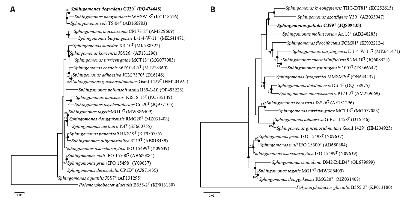
Fig. 2.Phylogenomic tree of two novel species and type strains of the genus Sphingomonas based on 81 UBCGs. This tree is a part of the phylogenomic tree including the entire species of Sphingomonas. Bootstrap values greater than 70% are shown at branch points based on maximum-likelihood analysis of 1,000 replicated datasets. Polymorphobacter B555-2T was used as an outgroup. Bar, 0.05 substitutions per nucleotide position.
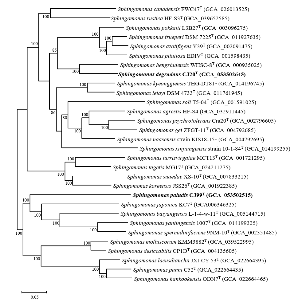
Table 1.
Genomic features of strains CJ20T, CJ99T and related type strains
Strains: 1, S. degradans CJ20T; 2, S. hengshuiensis WHSC-8T; 3, S. pokkalii L3B27TT; 4, S. azotifigens Y39T; 5, S. pituitosa EDIVT; 6, S. paludis CJ99T; 7, S. japonica KC7T; 8, S. baiyangensis L-1-4-w-11T; 9, S. yantingensis 1007T.
|
Genome feature |
1 |
2 |
3 |
4 |
5 |
6 |
7 |
8 |
9 |
|
Size (Mb) |
4.63 |
4.8 |
4.2 |
5.1 |
4.7 |
3.17 |
2.9 |
3.3 |
3.6 |
|
G + C (%) |
67.0 |
66.7 |
66 |
67.5 |
67 |
65.8 |
66.5 |
68.0 |
68.0 |
|
CDS |
4183 |
4784 |
3846 |
4656 |
4340 |
2991 |
2784 |
3153 |
3430 |
|
Contigs |
2 |
2 |
34 |
163 |
75 |
1 |
1 |
7 |
11 |
|
rRNA genes |
6 |
7 |
3 |
3 |
5 |
3 |
3 |
3 |
3 |
|
tRNA genes |
52 |
49 |
48 |
44 |
46 |
46 |
46 |
47 |
47 |
Table 2.
ANI and dDDH values between strains CJ20T, CJ99T and related type strains
Strains: 1, S. degradans CJ20T; 2, S. hengshuiensis WHSC-8T; 3, S. pokkalii L3B27TT; 4, S. azotifigens Y39T; 5, S. pituitosa EDIVT; 6, S. paludis CJ99T; 7, S. japonica KC7T; 8, S. baiyangensis L-1-4-w-11T; 9, S. yantingensis 1007T.
|
dDDH |
1 |
2 |
3 |
4 |
5 |
6 |
7 |
8 |
9 |
|
ANI |
|
1 |
|
34.4 |
20.9 |
22.0 |
22.3 |
16.1 |
17.4 |
17.5 |
17.3 |
|
2 |
82.9 |
|
19.5 |
20.5 |
20.6 |
15.5 |
17.2 |
17.8 |
17.1 |
|
3 |
78.1 |
77.3 |
|
40.2 |
36.9 |
15.0 |
16.4 |
16.6 |
16.6 |
|
4 |
78.3 |
77.7 |
85.1 |
|
43.4 |
14.8 |
16.5 |
16.7 |
16.9 |
|
5 |
78.4 |
78.0 |
84.7 |
87.1 |
|
15.1 |
16.6 |
17.1 |
17.3 |
|
6 |
74.4 |
74.0 |
73.7 |
73.9 |
73.9 |
|
16.8 |
16.1 |
16.2 |
|
7 |
75.3 |
75.5 |
74.5 |
75.0 |
74.9 |
74.3 |
|
23.8 |
20.7 |
|
8 |
75.3 |
75.3 |
74.6 |
75.2 |
74.9 |
74.3 |
76.8 |
|
25.2 |
|
9 |
75.0 |
75.0 |
74.2 |
74.8 |
74.8 |
74.1 |
76.2 |
77.9 |
|
Table 3.
Differential characteristics of strains CJ20T, CJ99T and related type strains of the genus Sphingomonas
Strains: 1, S. degradans CJ20T; 2, S. hengshuiensis WHSC-8T; 3, S. soli T5-04T; 4, S. paludis CJ99T; 5, S. kyeonggiensis THG-DT81T; 6, S. azotifigens Y39T; +, positive; −, negative.
|
Characteristics |
1 |
2 |
3 |
4 |
5 |
6 |
|
Isolation source* |
Han River |
Hengshui Lakea
|
Ginseng fieldb
|
Wetland |
Ginseng fieldc
|
Rootsd
|
|
Motility |
Non-motile |
Motilea
|
Non-motileb
|
Motile |
Motilec
|
Motiled
|
|
Colony color |
Yellow |
Yellow |
Yellow |
Yellow |
Yellow |
Yellow-Orange |
|
Growth
|
|
|
|
|
|
|
|
Temperature (°C) |
10–37 |
20–35a
|
15–37b
|
10–40 |
15–30c
|
25–37d
|
|
pH |
6.0–8.0 |
5.0–10.0a
|
6.8–7.5b
|
7.0–8.0 |
6.0–9.5c
|
ND |
|
NaCl (%, w/v) |
0–3 |
0–1a
|
ND |
0–2 |
0–0.5c
|
0–2.5d
|
|
Hydrolysis of
|
|
|
|
|
|
|
|
Starch |
− |
− |
− |
+ |
− |
− |
|
Cellulose |
− |
− |
− |
+ |
− |
− |
|
Casein |
− |
− |
− |
+ |
− |
− |
|
DNA |
− |
+ |
+ |
+ |
− |
+ |
|
Assimilation of
|
|
|
|
|
|
|
|
ᴅ-Mannose |
+ |
+ |
− |
+ |
+ |
+ |
|
ᴅ-Mannitol |
− |
+ |
− |
− |
− |
− |
|
N-Acetyl-glucosamine |
+ |
− |
− |
− |
+ |
+ |
|
ᴅ-Maltose |
+ |
+ |
− |
+ |
+ |
+ |
|
Potassium gluconate |
− |
+ |
− |
+ |
− |
− |
|
Adipic acid |
− |
+ |
− |
+ |
− |
− |
|
Malic acid |
+ |
+ |
− |
+ |
+ |
+ |
|
Enzyme activity
|
|
|
|
|
|
|
|
Oxidase |
− |
+ |
+ |
+ |
+ |
+ |
|
ᴅ-Glucose fermentation |
− |
− |
− |
− |
− |
+ |
|
β-Galactosidase |
+ |
+ |
+ |
+ |
− |
+ |
Table 4.
Fatty acid composition (%) of strains CJ20T, CJ99T and related type strains of genus Sphingomonas
Strains: 1, S. degradans CJ20T; 2, S. hengshuiensis WHSC-8T; 3, S. soli T5-04T; 4, S. paludis CJ99T; 5, S. kyeonggiensis THG-DT81T; 6, S. azotifigens Y39T; TR, trace amount (< 1%); –, not detected.
|
Fatty acid |
1 |
2 |
3 |
4 |
5 |
6 |
|
Saturated
|
|
|
|
|
|
|
|
iso–C10:0
|
− |
5.3 |
− |
1.6 |
9.0 |
5.0 |
|
C16:0
|
15.2
|
13.6
|
18.9
|
17.5
|
21.2
|
24.2
|
|
C17:0
|
− |
− |
− |
1.4 |
− |
− |
|
C18:0
|
− |
− |
− |
− |
1.2 |
1.4 |
|
Unsaturated
|
|
|
|
|
|
|
|
C16:1 ω5c
|
− |
− |
− |
1.4 |
− |
− |
|
C17:1 ω6c
|
− |
5.4 |
3.8 |
15.1 |
2.7 |
TR |
|
C17:1 ω8c
|
− |
− |
− |
2.5 |
− |
− |
|
C18:1 ω5c
|
− |
− |
− |
− |
− |
1.1 |
|
11methyl–C18:1 ω7c
|
− |
− |
4.6 |
2.7 |
4.3 |
5.9 |
|
cyclo–C19:0 ω8c
|
− |
− |
− |
− |
− |
TR |
|
Hydroxy
|
|
|
|
|
|
|
|
C8:0 3–OH |
5.5 |
− |
− |
− |
− |
− |
|
C14:0 2–OH |
3.7 |
7.2 |
− |
2.0 |
4.7 |
4.5 |
|
Summed features*
|
|
|
|
|
|
|
|
3 |
3.0 |
5.3 |
7.5 |
9.0 |
− |
− |
|
8 |
71.4
|
61.5
|
64.5
|
44.6
|
55.8
|
55.1
|
References
- Alcock BP, Huynh W, Chalil R, Smith KW, Raphenya AR, et al. 2023. Card 2023: expanded curation, support for machine learning, and resistome prediction at the comprehensive antibiotic resistance database. Nucleic Acids Res. 51: D690–D699. ArticlePubMedPMCPDF
- Asaf S, Numan M, Khan AL, Al-Harrasi A. 2020. Sphingomonas: From diversity and genomics to functional role in environmental remediation and plant growth. Crit Rev Biotechnol. 40: 138–152. ArticlePubMed
- Baker G, Smith JJ, Cowan DA. 2003. Review and re-analysis of domain-specific 16S primers. J Microbiol Methods. 55: 541–555. ArticlePubMed
- Bouras G, Houtak G, Wick RR, Mallawaarachchi V, Roach MJ, et al. 2024. Hybracter: enabling scalable, automated, complete and accurate bacterial genome assemblies. Microb Genom. 10: 001244.ArticlePubMedPMC
- Chen J, Wong MH, Wong YS, Tam NF. 2008. Multi-factors on biodegradation kinetics of polycyclic aromatic hydrocarbons (PAHs) by Sphingomonas sp. a bacterial strain isolated from mangrove sediment. Mar Pollut Bull. 57: 695–702. ArticlePubMed
- Choi I, Srinivasan S, Kim MK. 2024. Sphingomonas immobilis sp. nov., and Sphingomonas natans sp. nov. bacteria isolated from soil. Arch Microbiol. 206: 278.ArticlePubMedPDF
- Chun J, Oren A, Ventosa A, Christensen H, Arahal DR, et al. 2018. Proposed minimal standards for the use of genome data for the taxonomy of prokaryotes. Int J Syst Evol Microbiol. 68: 461–466. ArticlePubMed
- Collins MD. 1985. 11 analysis of isoprenoid quinones. In Bergan T. (ed.), Methods in microbiology, pp. 329–366. Elsevier. Article
- Dong L, Li S, Lian WH, Wei QC, Mohamad OAA, et al. 2022. Sphingomonas arenae sp. nov., isolated from desert soil. Int J Syst Evol Microbiol. 72: 005195.Article
- Dusa A. 2024. venn: Draw Venn Diagrams. R package version 1.12.2. Available from https://github.com/dusadrian/venn.
- Edgar RC. 2004. Muscle: Multiple sequence alignment with high accuracy and high throughput. Nucleic Acids Res. 32: 1792–1797. ArticlePubMedPMC
- Fujii K, Urano N, Ushio H, Satomi M, Kimura S. 2001. Sphingomonas cloacae sp. nov., a nonylphenol-degrading bacterium isolated from wastewater of a sewage-treatment plant in Tokyo. Int J Syst Evol Microbiol. 51: 603–610. ArticlePubMed
- Jukes TH, Cantor CR. 1969. Chapter 24 - Evolution of protein molecules. In Munro HN. (ed.), Mammalian protein metabolism, vol. 3rd, pp. 21–132. Academic Press. Article
- Kanehisa M, Sato Y, Morishima K. 2016. BlastKOALA and GhostKOALA: KEGG tools for functional characterization of genome and metagenome sequences. J Mol Biol. 428: 726–731. ArticlePubMed
- Kim SJ, Moon JY, Lim JM, Ahn JH, Weon HY, et al. 2014a. Sphingomonas aerophila sp. nov. and Sphingomonas naasensis sp. nov., isolated from air and soil, respectively. Int J Syst Evol Microbiol. 64: 926–932. Article
- Kim J, Na SI, Kim D, Chun J. 2021. UBCG2: Up-to-date bacterial core genes and pipeline for phylogenomic analysis. J Microbiol. 59: 609–615. ArticlePubMedPDF
- Kim M, Oh HS, Park SC, Chun J. 2014b. Towards a taxonomic coherence between average nucleotide identity and 16S rRNA gene sequence similarity for species demarcation of prokaryotes. Int J Syst Evol Microbiol. 64: 346–351. Article
- Kosako Y, Yabuuchi E, Naka T, Fujiwara N, Kobayashi K. 2000. Proposal of Sphingomonadaceae fam. nov., consisting of Sphingomonas Yabuuchi et al. 1990, Erythrobacter Shiba and Shimidu 1982, Erythromicrobium Yurkov et al. 1994, Porphyrobacter Fuerst et al. 1993, Zymomonas Kluyver and van Niel 1936, and Sandaracinobacter Yurkov et al. 1997, with the type genus Sphingomonas Yabuuchi et al. 1990. Microbiol Immunol. 44: 563–575. ArticlePubMed
- Lee I, Kim YO, Park SC, Chun J. 2016. OrthoANI: an improved algorithm and software for calculating average nucleotide identity. Int J Syst Evol Microbiol. 66: 1100–1103. ArticlePubMed
- Manni M, Berkeley MR, Seppey M, Zdobnov EM. 2021. BUSCO: assessing genomic data quality and beyond. Curr Protoc. 1: e323. ArticlePubMedLink
- Meier-Kolthoff JP, Carbasse JS, Peinado-Olarte RL, Göker M. 2022. TYGS and Lpsn: a database tandem for fast and reliable genome-based classification and nomenclature of prokaryotes. Nucleic Acids Res. 50: D801–D807. ArticlePubMedPMCPDF
- Minh BQ, Schmidt HA, Chernomor O, Schrempf D, Woodhams MD, et al. 2020. IQ-TREE 2: New models and efficient methods for phylogenetic inference in the genomic era. Mol Biol Evol. 37: 1530–1534. ArticlePubMedPMCPDF
- Minnikin D, O'donnell A, Goodfellow M, Alderson G, Athalye M, et al. 1984. An integrated procedure for the extraction of bacterial isoprenoid quinones and polar lipids. J Microbiol Methods. 2: 233–241. Article
- Parte AC, Sardà Carbasse J, Meier-Kolthoff JP, Reimer LC, Göker M. 2020. List of prokaryotic names with standing in nomenclature (LPSN) moves to the DSMZ. Int J Syst Evol Microbiol. 70: 5607–5612. ArticlePubMedPMC
- Richter M, Rosselló-Móra R. 2009. Shifting the genomic gold standard for the prokaryotic species definition. Proc Natl Acad Sci USA. 106: 19126–19131. ArticlePubMedPMC
- Romanenko LA, Tanaka N, Frolova GM, Mikhailov VV. 2009. Sphingomonas japonica sp. nov., isolated from the marine crustacean Paralithodes camtschatica. Int J Syst Evol Microbiol. 59: 1179–1182. ArticlePubMed
- Rosselló-Móra R, Amann R. 2015. Past and future species definitions for Bacteria and Archaea. Syst Appl Microbiol. 38: 209–216. ArticlePubMed
- Sasser M. 1990. Identification of bacteria by gas chromatography of cellular fatty acids. MIDI technical note 101. MIDI Inc.PDF
- Sayers EW, Bolton EE, Brister JR, Canese K, Chan J, et al. 2022. Database resources of the national center for biotechnology information. Nucleic Acids Res. 50: D20–D26. ArticlePubMed
- Schwengers O, Jelonek L, Dieckmann MA, Beyvers S, Blom J, et al. 2021. Bakta: rapid and standardized annotation of bacterial genomes via alignment-free sequence identification. Microb Genom. 7: 000685.ArticlePubMedPMC
- Son HM, Kook M, Tran HT, Kim KY, Park SY, et al. 2014. Sphingomonas kyeonggiense sp. nov., isolated from soil of a ginseng field. Antonie van Leeuwenhoek. 105: 791–797. ArticlePubMedPDF
- Srivathsan A, Meier R. 2012. On the inappropriate use of Kimura‐2‐parameter (K2P) divergences in the DNA‐barcoding literature. Cladistics. 28: 190–194. ArticlePubMed
- Takeuchi M, Hamana K, Hiraishi A. 2001. Proposal of the genus Sphingomonas sensu stricto and three new genera, Sphingobium, Novosphingobium and Sphingopyxis, on the basis of phylogenetic and chemotaxonomic analyses. Int J Syst Evol Microbiol. 51: 1405–1417. ArticlePubMed
- Tamura K, Stecher G, Kumar S. 2021. MEGA11: molecular evolutionary genetics analysis version 11. Mol Biol Evol. 38: 3022–3027. ArticlePubMedPMCPDF
- Tonkin-Hill G, MacAlasdair N, Ruis C, Weimann A, Horesh G, et al. 2020. Producing polished prokaryotic pangenomes with the Panaroo pipeline. Genome Biol. 21: 180.ArticlePubMedPMCPDF
- Wang Z, Zeng Q, Fang Z, Zhu D, Xu D, et al. 2019. Sphingomonas aracearum sp. nov., isolated from rhizospheric soil of Araceae plants. Int J Syst Evol Microbiol. 69: 2972–2978. ArticlePubMed
- Wei S, Wang T, Liu H, Zhang C, Guo J, et al. 2015. Sphingomonas hengshuiensis sp. nov., isolated from lake wetland. Int J Syst Evol Microbiol. 65: 4644–4649. ArticlePubMed
- Wick RR, Judd LM, Gorrie CL, Holt KE. 2017. Completing bacterial genome assemblies with multiplex MinION sequencing. Microb Genom. 3: e000132. ArticlePubMedPMC
- Xie CH, Yokota A. 2006. Sphingomonas azotifigens sp. nov., a nitrogen-fixing bacterium isolated from the roots of Oryza sativa. Int J Syst Evol Microbiol. 56: 889–893. ArticlePubMed
- Yabuuchi E, Yano I, Oyaizu H, Hashimoto Y, Ezaki T, et al. 1990. Proposals of Sphingomonas paucimobilis gen. nov. and comb. nov., Sphingomonas parapaucimobilis sp. nov., Sphingomonas yanoikuyae sp. nov., Sphingomonas adhaesiva sp. nov., Sphingomonas capsulata comb, nov., and two genospecies of the genus Sphingomonas. Microbiol Immunol. 34: 99–119. ArticlePubMed
- Yang DC, Im WT, Kim MK, Ohta H, Lee ST. 2006. Sphingomonas soli sp. nov., a β-glucosidase-producing bacterium in the family Sphingomonadaceae in the α-4 subgroup of the Proteobacteria. Int J Syst Evol Microbiol. 56: 703–707. ArticlePubMed
- Yoon SH, Ha SM, Kwon S, Lim J, Kim Y, et al. 2017. Introducing EzBioCloud: a taxonomically united database of 16S rRNA gene sequences and whole-genome assemblies. Int J Syst Evol Microbiol. 67: 1613–1617. ArticlePubMedPMC
- Yoon JH, Park S, Kang SJ, Kim W, Oh TK. 2009. Sphingomonas hankookensis sp. nov., isolated from wastewater. Int J Syst Evol Microbiol. 59: 2788–2793. ArticlePubMed
- Zhou Z, Tran PQ, Breister AM, Liu Y, Kieft K, et al. 2022. METABOLIC: high-throughput profiling of microbial genomes for functional traits, metabolism, biogeochemistry, and community-scale functional networks. Microbiome. 10: 33.ArticlePubMedPMCPDF
Citations
Citations to this article as recorded by


 ePub Link
 Cite this Article


